
Angiomas and Vascular Anomalies Unit
At our clinics in Madrid: Calle Serrano 143 and La Moraleja
Dermatologists specialising in
Treatment of angiomas and other vascular anomalies
Angiomas are benign vascular anomalies that affect approximately one in ten newborn babies. Although their cause is unknown and they can’t be prevented, they often disappear on their own in early childhood. The Grupo Pedro Jaén angioma experts recommend diagnosis and monitoring of patients in order to treat them if there is aesthetic damage and, most importantly, to differentiate angiomas from vascular malformations. Vascular malformations are a more serious clinical condition that may require early treatment, a decision that is taken after referring the patient to our Vascular Malformations Unit.

Recognised reference clinics in Spain
Home > Clinical Dermatology > Angiomas and Vascular Anomalies
What sets us apart
Multidisciplinary approach tailored to each case
Our Angioma and Vascular Anomalies Unit offers a highly specialised approach to the diagnosis and treatment of these benign vascular skin lesions, combining clinical precision with advanced technology. We use Doppler ultrasound and specific markers such as GLUT-1 to differentiate between haemangiomas and vascular malformations, avoiding inappropriate treatments and ensuring the most appropriate therapeutic approach. We have the most comprehensive laser platform in Europe for treating visible traces left on the skin and work closely with vascular and paediatric surgery in complex cases. Our multidisciplinary and personalised approach enables us to offer safe, effective and personalised solutions for each patient.
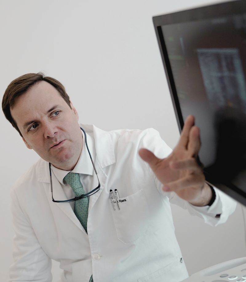
Advanced diagnostics
Accurate diagnosis is key to designing effective treatment and avoiding complications when treating vascular anomalies. Our unit uses state-of-the-art technology to accurately identify each type of vascular mark and determine the best therapeutic approach.
Doppler ultrasound and specific biomarkers
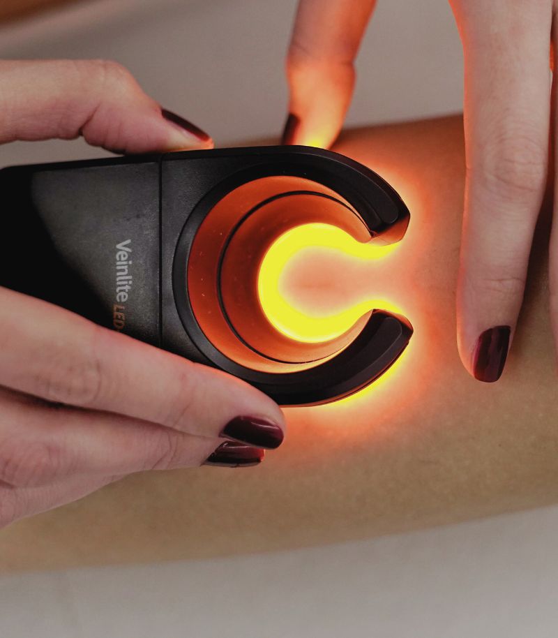
Our specialists in angiomas and other vascular anomalies use high-resolution Doppler ultrasound to assess blood flow and differentiate between infantile haemangiomas, vascular malformations and other vascular anomalies. In addition, we use biomarkers such as GLUT-1, which are essential to distinguish haemangiomas from other abnormalities that may require completely different treatment strategies.
Complementary examinations for a complete assessment
In complex cases, MRI, CT and angiographic studies are used to determine the extent of the vascular anomaly and its relationship with nearby structures. These tests enable more precise therapeutic planning and help predict the evolution of the anomaly, reducing the risk of functional or aesthetic complications. Our detailed approach ensures that each patient receives an accurate diagnosis and treatment tailored to their clinical situation.
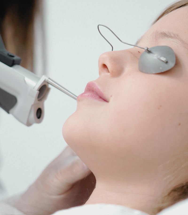
Specific therapies for each vascular anomaly
Each vascular anomaly requires a tailored treatment, combining medical therapies, laser therapy and minimally invasive procedures to achieve the best results with maximum safety.
Personalised pharmacological therapies
For infantile haemangiomas and superficial anomalies, we apply topical, oral and injectable treatments that stop them growing and promote gradual shrinking. These treatments reduce the need for invasive procedures in many cases and minimise the aesthetic impact of the affected area.
Vascular laser for persistent skin anomalies
Pulsed dye lasers and other advanced technologies are highly effective in treating involuted angiomas, cherry angiomas and telangiectasias. These techniques offer visible results from the first sessions, with minimal recovery times and no scarring. In addition, laser treatment is a safe and effective option even on delicate areas such as the face or neck, resulting in more even skin and a better patient experience.
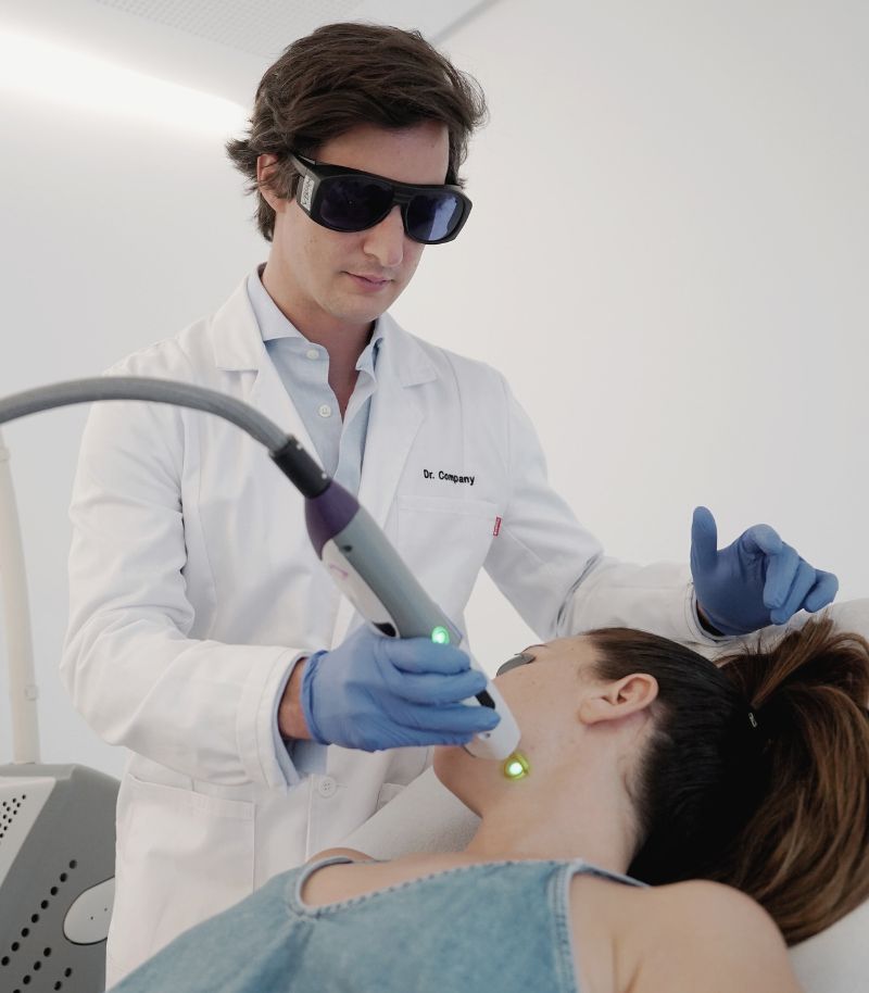
Approach to complex vascular malformations
Some complex vascular alterations—such as larger vascular malformations, superficial venous anomalies (including venous ectasias and varicose veins), as well as benign vascular tumours—are referred to our Vascular Malformations Unit, which in turn is supported by the Vascular Surgery Unit.
Vascular Malformations Unit
This team deals with congenital and acquired vascular anomalies through a multidisciplinary and cross-functional approach involving other units of the group, such as the Laser Unit. They also have the support of our team of dermatologists and an external plastic surgeon who specialises in vascular malformations. Patients can be referred to this surgeon after the initial assessment.
Integrated care protocol
The Grupo Pedro Jaén specialists who treat vascular malformations have established a protocol which streamlines the healthcare process that supports patients throughout their treatment. This reduces the time that elapses from the moment they receive the diagnosis until they access treatment, as well as avoiding opinions which are sometimes contradictory.
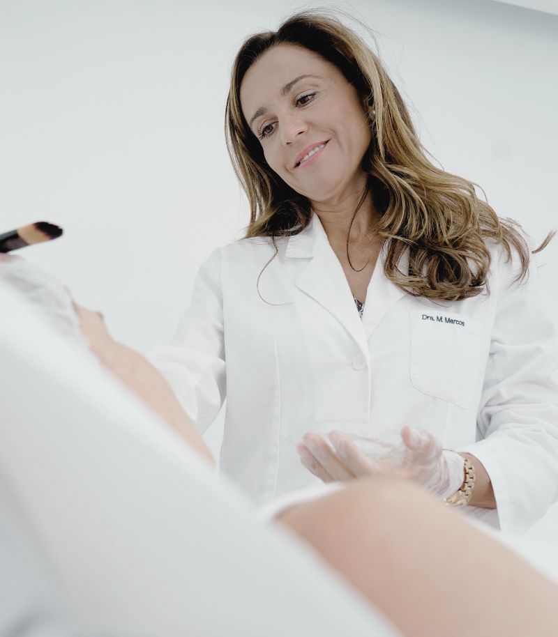
Cross-functional work with other units in the group
The treatment of angiomas covers vascular anomalies and malformations of varying severity, such as haemangiomas, strawberry birthmarks, cherry angiomas, venous lakes, venous malformations, telangiectasias, varicose veins and cuperosis.
Multidisciplinary team
Treating angiomas may require dermatologists, vascular surgeons, plastic surgeons, diagnostic imaging experts, paediatric surgeons and rehabilitation and make-up specialists. Grupo Pedro Jaén has specific units and external collaborators who can offer comprehensive care to each patient.
Laser and Aesthetic Rehabilitation Unit
Our Laser Unit is one of the most advanced in Europe and can complement vascular treatments with specific technologies to eliminate visible marks while respecting the skin. This minimises the aesthetic and functional consequences for patients whose vascular anomalies have left marks or thickened skin. In addition, we integrate aesthetic rehabilitation techniques to promote skin recovery and optimise treatment results.
Treatments for sensitive areas
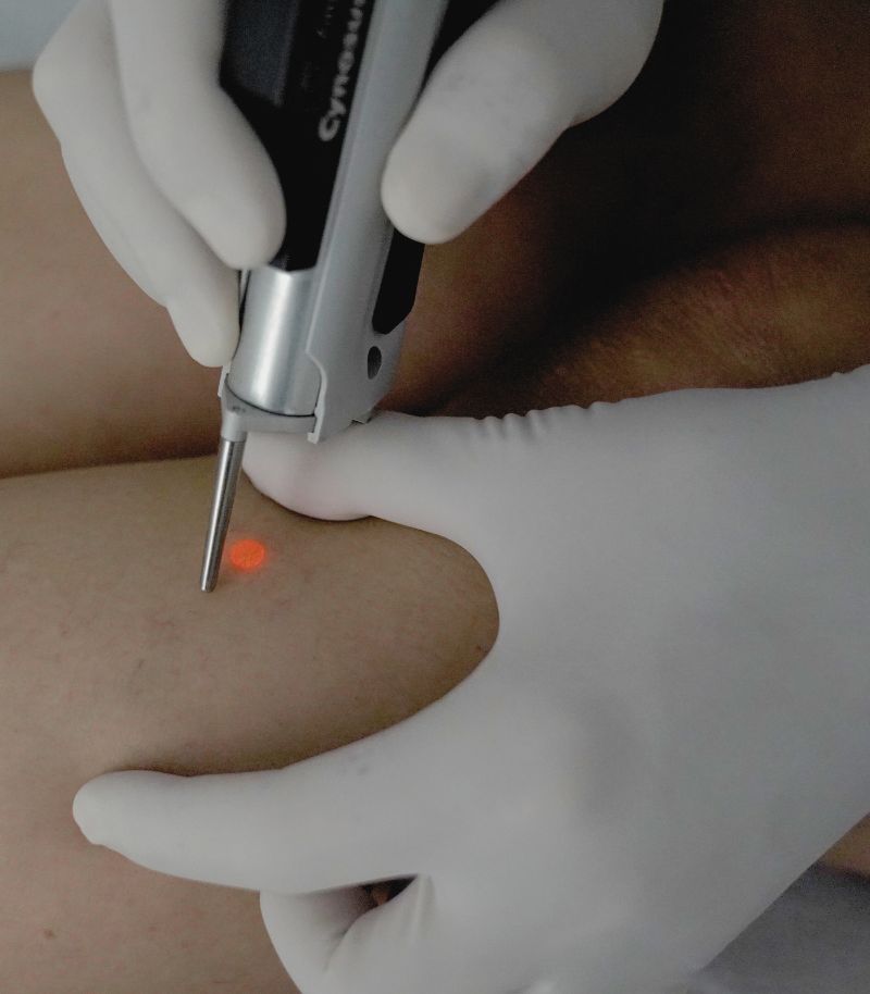
Vascular anomalies in sensitive areas such as the lips, face and other exposed areas require a precise approach to ensure removal of the anomaly without compromising the skin’s appearance and functionality.
Nd:YAG laser for deeper vascular anomalies
The Nd:YAG laser is the most effective treatment option for venous lakes and other deeper vascular anomalies. Its selective coagulation capability enables the removal of abnormalities without damaging the surrounding tissues, ensuring rapid recovery and no visible scarring. Thanks to its precision, it has become the treatment of choice for anomalies in delicate areas such as the lips and face, achieving safe and lasting results.
Combining advanced techniques for optimal results
In more complex cases, we combine different strategies such as cryotherapy, sclerotherapy or minimally invasive surgery to enhance the effectiveness of the treatment. This combination enables the procedure to be adapted to the individual characteristics of the patient, achieving both functional and aesthetic improvement. With this personalised approach, we guarantee maximum safety and efficacy in the management of specific vascular anomalies.
Innovation and development
Our commitment to research and technology enables us to offer cutting-edge treatments for angiomas and vascular malformations, optimising clinical and aesthetic results.
Participation in international clinical studies
Our specialists are global leaders in their field and are involved in cutting-edge research to develop new therapeutic strategies to treat complex vascular anomalies. Thanks to these research projects, we contribute to the evolution of safer, more effective and personalised treatments, ensuring that our patients have access to innovative options backed by scientific evidence.
Evolving therapeutic strategies
The continuous enhancement of our experts’ knowledge enables us to incorporate the most advanced techniques in the management of angiomas and vascular malformations. We regularly update our protocols guided by the latest consensus in vascular dermatology, ensuring more precise treatments, reduced side effects and lasting results. This approach establishes us as recognised leaders in delivering innovative treatments for vascular anomalies.
What they are and the different types
A guide to understanding angiomas
The term haemangioma refers to benign vascular lesions that grow rapidly during early infancy (within the first year of life), characterised by a phase of rapid proliferation followed by gradual involution. Early assessment of each case is essential in order to determine whether treatment is required and, if so, to apply it at the most appropriate time to achieve the best possible outcome.
Traditionally, however, the term angioma has also been used to describe other vascular lesions that are amenable to laser treatment, including:
- Strawberry birthmarks.
- Venous lakes.
- Cherry angiomas.
- Senile angiomas.
- Telangiectasias.

Haemangiomas (“strawberry birthmarks”)
This type of angioma or haemangioma is relatively common in childhood. They are popularly known as strawberry birthmarks because they appear as bright red lesions formed by clusters of abnormally proliferating blood vessels that are visible on the surface of the skin.
They most commonly affect the nose, mouth, vagina or anus, although they can appear on any part of the body. They tend to be slightly raised, have a soft texture and are compressible to the touch.
The reported incidence of haemangiomas varies widely, but it is generally estimated that they affect around 1–3% of newborns, rising to up to 12% in infants under one year of age. Around one third of these benign tumours are diagnosed at birth and are therefore often classified as birthmarks.
However, it is estimated that only around 20% of infantile angiomas are present at birth, while the remaining 80% develop between two and four weeks of life.
Causes of haemangiomas
The exact causes of the abnormal vascular proliferation that gives rise to infantile haemangiomas are not fully understood. What is known is that these angiomas or strawberry birthmarks are much more common in girls and, according to medical literature, may follow an autosomal dominant inheritance pattern. This means that if one parent carries the gene or genes associated with the anomaly, the child has a 50% chance of inheriting it.
Premature babies and those born with low birth weight are also more likely to develop angiomas than infants born at term with a normal weight.
It has also been demonstrated that strawberry birthmarks express a marker known as GLUT-1, a glucose transporter found in the blood–brain barrier and in red blood cells.
This marker provides specialists with highly valuable information when differentiating angiomas from other types of vascular malformations, which is a key factor in selecting the most appropriate treatment. Although most infantile haemangiomas are reddish in colour, some may appear bluish. These tend to be located deeper in the skin and require thorough assessment to determine whether they are benign tumours or another type of vascular malformation with a less favourable prognosis.
Evolution of infantile haemangiomas
Strawberry birthmarks follow a very similar course in most patients. Before becoming visible, a faint pink, bluish or whitish patch may appear, often going unnoticed. In the next phase, the angioma becomes raised and gradually acquires its characteristic red colour.
After a period of rapid vascular proliferation, growth and maturation, the infantile haemangioma enters a regression phase. Growth stops and the lesion gradually fades. Around 50% resolve by the age of five, 70% by the age of seven, and over 90% leave no visible trace by the age of nine. If any residue remains, it is usually mild, such as a telangiectasia or redundant skin.
How haemangiomas are diagnosed
In addition to clinical examination and assessment of the patient’s medical and family history, infantile haemangiomas are diagnosed using Doppler ultrasound or, when necessary, MRI or analysis of the GLUT-1 marker.
The aim of these tests is to differentiate strawberry birthmarks from other vascular malformations or tumours and to obtain detailed information regarding their location, extent, aesthetic or functional implications and likely evolution.
Treatment of infantile haemangiomas
In most cases, infantile angiomas do not require specific treatment, as they are benign lesions that tend to resolve spontaneously over time.
Nevertheless, close monitoring is essential to confirm favourable progression, detect early changes and treat complications should they arise. Although angiomas are benign, ulceration can occur in approximately 13% of cases, particularly when lesions are located in areas subject to friction, such as the buttocks.
As a result, a watchful waiting approach is usually adopted, with clinical intervention reserved for cases at high risk of ulceration and subsequent scarring or deformity. At the Grupo Pedro Jaén Vascular Anomalies Unit, each case is assessed individually to determine the most appropriate management strategy.
When medical treatment is required, earlier intervention is associated with a better response and outcome, highlighting the importance of early specialist assessment.
Medications for haemangiomas
Different types of medication may be used depending on the size, location and characteristics of the lesion.
Oral or topical treatment
These therapies help to halt haemangioma growth and promote regression. Topical formulations are particularly useful for small, superficial lesions at an early stage, provided they are not located on mucous membranes and are not ulcerated.
Localised injectable treatment
Medications can be injected directly into the haemangioma to achieve highly localised size reduction. This approach is especially suitable for well-defined cutaneous haemangiomas located on the ear, nasal tip, cheeks, eyelids or lips and may be combined with oral therapy in selected cases.
Other pharmacological options
Additional therapies may be used in complicated haemangiomas to modulate the immune system, inhibit angiogenesis (formation of new blood vessels) or induce apoptosis (programmed cell death), thereby controlling the vascular component of the lesion.
Laser treatment for haemangiomas
Vascular laser therapy offers excellent results in involuted (residual), abortive and ulcerated haemangiomas, promoting rapid re-epithelialisation and lesion closure. In some cases, it may be combined with beta-blockers depending on the patient.
Vascular lasers can also be combined with other laser modalities to treat skin thickening, improve scarring (ablative and non-ablative fractional lasers) and reduce residual sequelae.
For this type of treatment, the Grupo Pedro Jaén Vascular Anomalies Unit works closely with the Laser Unit, the largest private laser platform in Europe, which collaborates transversally with all medical specialties within the group.
Surgery for infantile haemangiomas
Although strawberry birthmarks generally have an excellent prognosis, surgery may be required in exceptional cases involving serious complications, such as recurrent bleeding, involvement or deformation of adjacent structures, or impairment of vital functions such as vision.
Surgical cases are referred to the Grupo Pedro Jaén Plastic Surgery Unit, where the most appropriate technique is selected on an individual basis according to the patient’s circumstances and the size and location of the lesion.
“Early assessment of each case is essential in order to determine the most appropriate treatment and apply it at the optimal time to achieve the best possible outcome”
Cherry angiomas
Commonly known as red moles or blood moles, cherry angiomas are actually small angiomas caused by dilation of capillaries that become visible on the surface of the skin.
What are cherry angiomas?
Also known as cherry angiomas or microangiomas, these benign vascular lesions usually appear on the trunk and upper limbs. They have a strong hereditary and hormonal component and do not develop in childhood, but rather increase with age, which is why they are also referred to as senile angiomas.
They are more common in individuals with fair skin, although other phototypes may also be affected. They are usually slightly raised and rarely exceed one or two millimetres in diameter, although larger lesions can occur.
The exact cause is not fully understood, but cherry angiomas appear more frequently in very fair-skinned individuals with inadequate sun protection. Hormonal changes during pregnancy have also been associated with their development, although they usually regress after delivery.
Diagnosis is primarily clinical, although digital dermoscopy may be used to exclude malignancy and confirm the diagnosis by revealing subtle structural features not visible to the naked eye.
Treatment of cherry angiomas
As cherry angiomas are benign, treatment is not required unless they pose an aesthetic concern or bleed frequently due to friction or minor trauma.
When treatment is indicated, vascular laser therapy is the treatment of choice. As with strawberry birthmarks, the laser selectively targets red-pigmented vascular structures without damaging surrounding tissue. The procedure is quick, virtually painless and requires no downtime.
Laser treatment for cherry angiomas is neither uncomfortable nor painful and does not require downtime or time off work.
Although cherry angiomas are benign and not moles, monitoring is recommended if lesions grow or develop irregular borders, as this may indicate a different diagnosis requiring specialist assessment.
Venous lakes
Venous lakes are another type of angioma treated at the Grupo Pedro Jaén Angioma Unit. They appear as bluish or violaceous (sometimes almost black), soft, raised lesions resembling a small sac filled with dark blood.
They most commonly affect the lips but may also appear on the face or ears. They usually measure less than five millimetres in diameter, grow slowly and are asymptomatic.
They typically occur in individuals over 50 years of age, particularly those with fair skin, although they can occasionally appear at younger ages.
Causes of venous lakes
Venous lakes are mainly associated with chronic sun exposure. Less commonly, they may result from trauma or genetic syndromes, in which case they are classified as vascular malformations rather than angiomas.
Treatment of venous lakes
Venous lakes do not require treatment for medical reasons, only for aesthetic concerns or when larger lesions are prone to bleeding.
Treatment options include cryotherapy, sclerotherapy or surgical excision. However, the most effective and safest option is vascular laser therapy using a Nd:YAG laser, which has a longer wavelength capable of coagulating deeper lesions.
In some cases, a combination of vascular lasers may be required to optimise results. Treatment is virtually painless and usually consists of one main session and a follow-up session to refine the outcome.
This device uses a longer wavelength that coagulates the lesion, which is located at a deeper level than other types of angiomas. In some cases, it may be necessary to combine two vascular lasers to achieve a more satisfactory result. In all cases, the treatment is virtually painless and typically involves one treatment session, with a second session to refine the outcome if required.
The Grupo Pedro Jaén Laser Unit offers a wide range of laser systems, allowing the most appropriate combination to be selected for each lesion.

More information
Below you will find the frequently asked questions, authorship, and references for the information provided on this page:
Frequently asked questions about angiomas
What is the difference between a haemangioma and a vascular malformation?
Because haemangiomas are vascular anomalies, terminology is often used in a confusing way for patients. Essentially, haemangiomas are benign vascular tumours of an intense red colour that appear a few days after birth. They grow during the first year of life and may later decrease in size or even disappear completely.
By contrast, vascular malformations are present from birth. Over time, they tend to become darker and hypertrophic (thickened in appearance) and never resolve spontaneously.
Can haemangiomas be prevented?
No. Haemangiomas are relatively common anomalies related to the immaturity of the vascular system. In fact, it is estimated that around 20% of premature babies develop an angioma. There is therefore no way to prevent their formation.
Which specialist should I see if I have a haemangioma?
Several specialists may be involved in the management of a haemangioma, depending mainly on its location and size. At the Grupo Pedro Jaén Angiomas Unit, each lesion is individually assessed to determine whether it is an angioma or a vascular malformation and to refer the patient to the most appropriate unit. In most cases, haemangiomas can be managed entirely within our Angiomas Unit and Laser Unit. However, some cases may require treatment within the Plastic Surgery Unit or the Vascular Malformations Unit.
Can a haemangioma reappear?
No, provided that the diagnosis is correct—haemangiomas are often initially confused with vascular malformations until the correct diagnosis is established—and that the treatment prescribed by the specialist is completed without premature interruption.
What is a port-wine stain?
Also known as flat angiomas, port-wine stains are vascular malformations present from birth. They are most commonly located on the face but may appear anywhere on the body. They are initially pink in colour and gradually darken over time, often becoming raised after the age of 20. One of the main advantages of laser treatment is its ability to act selectively on affected tissues without damaging healthy skin or altering surrounding structures.
The mechanism of action is based on the laser’s affinity for the red colour of haemoglobin. In this way, vascular lasers destroy abnormal blood vessels and reduce the size, colour and texture of the lesion over a variable number of treatment sessions.
Authorship and references
The content of this page has been prepared by the Communication Department and the Medical Team of Grupo Pedro Jaén, in line with our editorial commitment to ensuring the accuracy and up-to-date nature of the information provided.
References on angiomas:
1. DeHart A, Richter G. Hemangioma: Recent Advances. F1000Res. 2019 Nov 18;8:F1000 Faculty Rev-1926. doi: 10.12688/f1000research.20152.1. PMID: 31807282; PMCID: PMC6871355.
2. Buslach N, Foulad DP, Saedi N, Mesinkovska NA. Treatment Modalities for Cherry Angiomas: A Systematic Review. Dermatol Surg. 2020 Dec;46(12):1691-1697. doi: 10.1097/DSS.0000000000002791. PMID: 33252464.
3. Rodríguez Bandera AI, Sebaratnam DF, Wargon O, Wong LF. Infantile hemangioma. Part 1: Epidemiology, pathogenesis, clinical presentation and assessment. J Am Acad Dermatol. 2021 Dec;85(6):1379-1392. doi: 10.1016/j.jaad.2021.08.019. Epub 2021 Aug 19. PMID: 34419524.
4. Jesús Alberto Sabando Carranza, Izaro Almandoz Irigoyen, Aitziber Arrugaeta Zabaleta, Experiencia en el tratamiento de los puntos rubí o angioma capilar adquirido en una consulta de atención primaria, FMC – Formación Médica Continuada en Atención Primaria, Volume 28, Issue 7, 2021, Pages 397-398, ISSN 1134-2072, DOI: 10.1016/j.fmc.2020.06.005.
5. Dua Cebeci, Seide Karasel, Şirin Yaşar, Venous Lakes of the Lips Successfully Treated With a Sclerosing Agent 1% polidocanol: analysis of 25 report cases, International Journal of Surgery Case Reports, Volume 78, 2021, Pages 265-269, ISSN 2210-2612, DOI: 10.1016/j.ijscr.2020.12.057.
6. H.E. John, H.S. Phen, P.J. Mahaffey, Treatment of venous lesions of the lips and perioral area with a long-pulsed Nd:YAG laser, British Journal of Oral and Maxillofacial Surgery, Volume 54, Issue 4, 2016, Pages 376-378, ISSN 0266-4356, DOI: 10.1016/j.bjoms.2015.06.014.
7. Sebaratnam DF, Rodríguez Bandera AL, Wong LF, Wargon O. Infantile hemangioma. Part 2: Management. J Am Acad Dermatol. 2021 Dec;85(6):1395-1404. doi: 10.1016/j.jaad.2021.08.020. Epub 2021 Aug 19. PMID: 34419523.
8. Mariani LG, Ferreira LM, Rovaris DL, Bonamigo RR, Kiszewski AE. Infantile hemangiomas: risk factors for complications, recurrence and unaesthetic sequelae. An Bras Dermatol. 2022 Jan-Feb;97(1):37-44. doi: 10.1016/j.abd.2021.05.009. Epub 2021 Nov 27. PMID: 34848114; PMCID: PMC8799848.
Where we see patients?
Clinics in Madrid
Grupo Pedro Jaén is a medical group founded more than three decades ago with the aim of providing medical, surgical and aesthetic solutions related to skin health and appearance. Led by Dr Pedro Jaén, the group has a highly specialised medical team that covers every area of care with top-level specialists, carries out extensive training activities, is a leader in medical research, and operates several clinics in Madrid, from which it offers the highest standards of care and clinical excellence to all patients.


Calle Serrano 143
This centre is the main headquarters of Grupo Pedro Jaén. It provides diagnostic and treatment services in skin cancer, clinical and aesthetic dermatology, laser treatments and other related specialties.
Mon - Fri 09:00 - 21:00 / Sat: 09:00 - 14:00
Calle Serrano 143, El Viso neighbourhood, Chamartín district, 28006 Madrid, Spain

La Moraleja
Located in Plaza de La Moraleja, this exclusive centre allows Grupo Pedro Jaén to bring its medical and aesthetic services closer to patients across the entire northern area of Madrid.
Mon - Thu: 09:30 - 20:00 / Fri: 09:30 - 18:00
Calle de la Estafeta 8, ground floor, La Moraleja, 28109 Alcobendas, Madrid
Related units and pages
Laser Unit
An international reference in laser and intense pulsed light treatments, both in clinical and aesthetic dermatology.
Vascular Surgery
Advanced diagnostic tools and treatments designed to deliver effective approaches with the least invasive techniques possible.
Diagnostic Imaging
Cutting-edge and highly effective imaging technologies to achieve more accurate diagnoses while avoiding invasive procedures.
Paediatric dermatology
Specialised care for the full range of dermatological conditions that may affect children.
Birthmarks
Each birthmark is carefully classified, a differential diagnosis is established and an appropriate management plan is designed.
Clinical dermatology
Highly specialised dermatologists for each skin condition, offering the most advanced treatments available.
Information and news
Press room
In the Grupo Pedro Jaén Press Room you can find information, advice and news related to dermatology, aesthetic medicine, trichology and general health. The group’s journals are also shared through an online platform, along with updates on the activity and media appearances of our clinics and healthcare professionals.

How to shower properly: tips for looking after your skin
Cold or hot water? Is it better to use a sponge? Find out how to shower properly and what dermatologists recommend to protect your skin.
Retinol in summer: should you stop using it?
We debunk this and other myths about using retinol during the warmer months.

Blog
News about Grupo Pedro Jaén. Information and advice on dermatology, aesthetic medicine, trichology, and general health

Media
Information about media appearances by our clinics and medical specialists.

Journal
Archive of all Grupo Pedro Jaén journals, available through an external online platform.