
Basal Cell Carcinoma
Treatment available at our Madrid clinics at Calle Serrano 143 and La Moraleja
Dermatologists specialising in
Treatment of basal cell carcinoma
Basal cell carcinoma is the most common type of skin tumour. Between 80% and 90% of all diagnosed cases of skin cancer are classified as basal cell carcinoma, also known as basalioma or basal cell epithelioma. It has a very limited ability to metastasise and usually grows slowly. However, early detection and treatment are essential, as the tumour progresses over time and may become locally invasive (in situ), potentially leading to aesthetic and functional damage.

Recognised reference clinics in Spain
Home > Skin Cancer > Basal Cell Carcinoma
What sets us apart
Effective treatment with aesthetic and functional preservation
Our Skin Cancer Unit is distinguished by its advanced approach to the diagnosis and treatment of basal cell carcinoma. We use state-of-the-art technology that allows lesions to be detected at very early stages, helping to avoid more aggressive treatments. Aesthetic and functional preservation are prioritised, and techniques such as Mohs surgery or photodynamic therapy are applied according to the characteristics of each case.
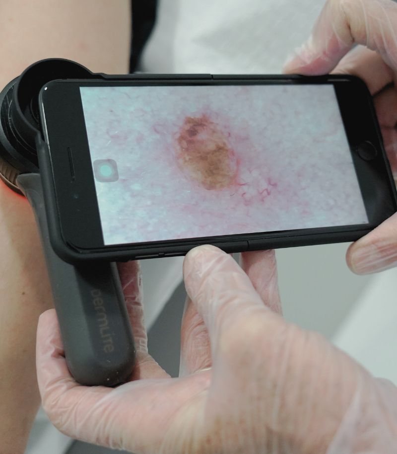
Early diagnosis
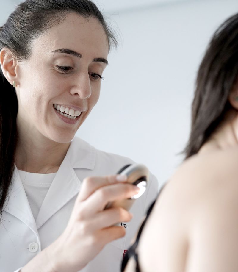
Early detection of basal cell carcinoma is key to achieving effective treatment with minimal impact. Our unit combines advanced diagnostic tools, including Vectra 360, confocal microscopy and digital dermoscopy, to ensure an accurate diagnosis. These technologies allow detailed analysis of tumour structures without the need for invasive procedures, facilitating early detection and improving cure rates.
Benefits of early diagnosis
Identifying the tumour at an early stage makes it possible to treat it before it grows or infiltrates healthy tissue, thereby avoiding extensive surgery or more aggressive procedures. This is particularly important in visible areas such as the face and neck, where aesthetic preservation is a priority.
Accuracy and safety in diagnosis
High-resolution diagnostic technology enables precise differentiation of basal cell carcinoma from other dermatological conditions. This ensures a tailored treatment strategy for each patient and optimises clinical outcomes, while avoiding unnecessary biopsies whenever possible.
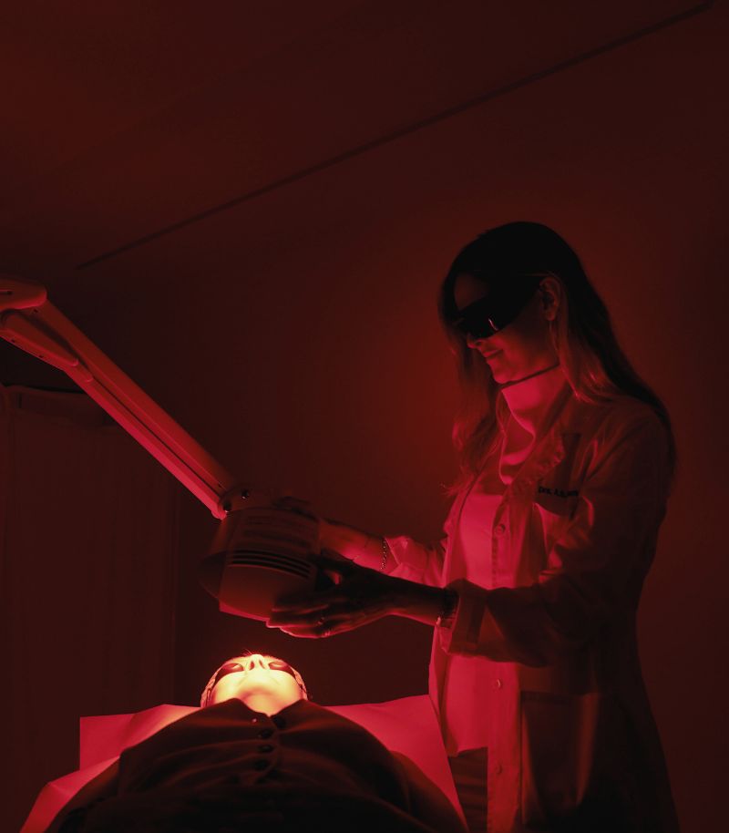
Aesthetic care
Basal cell carcinoma often appears in highly visible areas of the body, such as the nose, chin, face, neck, décolletage, ears or upper back. This can have a significant impact on a patient’s appearance and represents a therapeutic challenge for specialist dermatologists, whose aim is to completely remove the tumour while preserving both function and aesthetic outcomes.
Mohs surgery
Mohs surgery is a highly effective surgical technique that fulfils three key objectives: complete tumour removal, preservation of function and maximal conservation of healthy tissue. This approach is particularly important when tissue reconstruction is required after tumour excision and is of special relevance given that most skin cancers are located on the face.
Photodynamic therapy
Photodynamic therapy (PDT) is a treatment option for superficial basal cell carcinoma and selected small nodular lesions, particularly when surgery can be avoided and aesthetic outcomes are a priority.
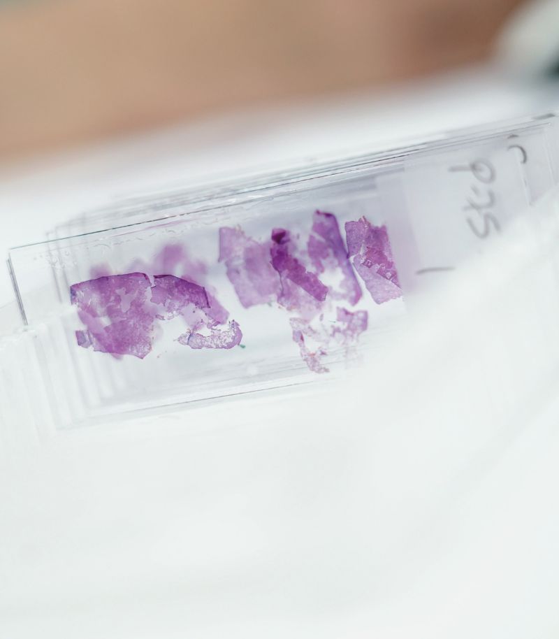
Personalised treatment
Skin cancer includes a wide range of tumours with different histological characteristics, prognoses and clinical considerations. For this reason, treatment decisions must always be made on an individual basis, taking multiple factors into account.
Tumour classification
Treatment planning is based on tumour location, size, depth of invasion, vascular involvement and whether the lesion has developed on pre-existing skin damage. These criteria allow tumours to be classified as low-risk or high-risk, guiding therapeutic decision-making.
Treatment tailored to each patient
Specialists in our Skin Cancer Unit take into account each patient’s age, life expectancy, clinical condition, risk factors and aesthetic considerations when selecting the most appropriate treatment. Potential complications and possible functional or aesthetic consequences are also carefully evaluated.
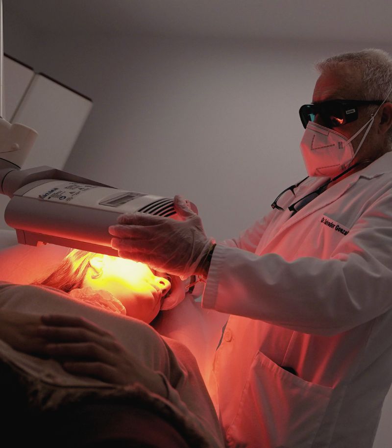
Innovation in non-invasive approaches
Non-invasive treatments have transformed the management of basal cell carcinoma, particularly in superficial lesions or in patients for whom less aggressive therapeutic options are appropriate.
Topical chemotherapy
Topical chemotherapy involves the application of a cream or gel directly to the cancerous lesion to destroy tumour cells. It is used to treat premalignant lesions and superficial basal cell carcinomas. Its highly localised action reduces the risk of systemic side effects and makes it a suitable option for lesions in visible areas such as the face and neck.
Photodynamic therapy
Photodynamic therapy is a form of photochemotherapy used to treat certain skin cancers in a non-invasive manner. The treatment involves applying a photosensitising cream to the affected area and exposing it to a specific light source, triggering a photochemical reaction that selectively destroys diseased cells while preserving healthy tissue.
Multidisciplinary approach
The Grupo Pedro Jaén Skin Cancer Unit works closely with other specialised units within the group to optimise treatment outcomes for basal cell carcinoma and to support recovery following surgery.
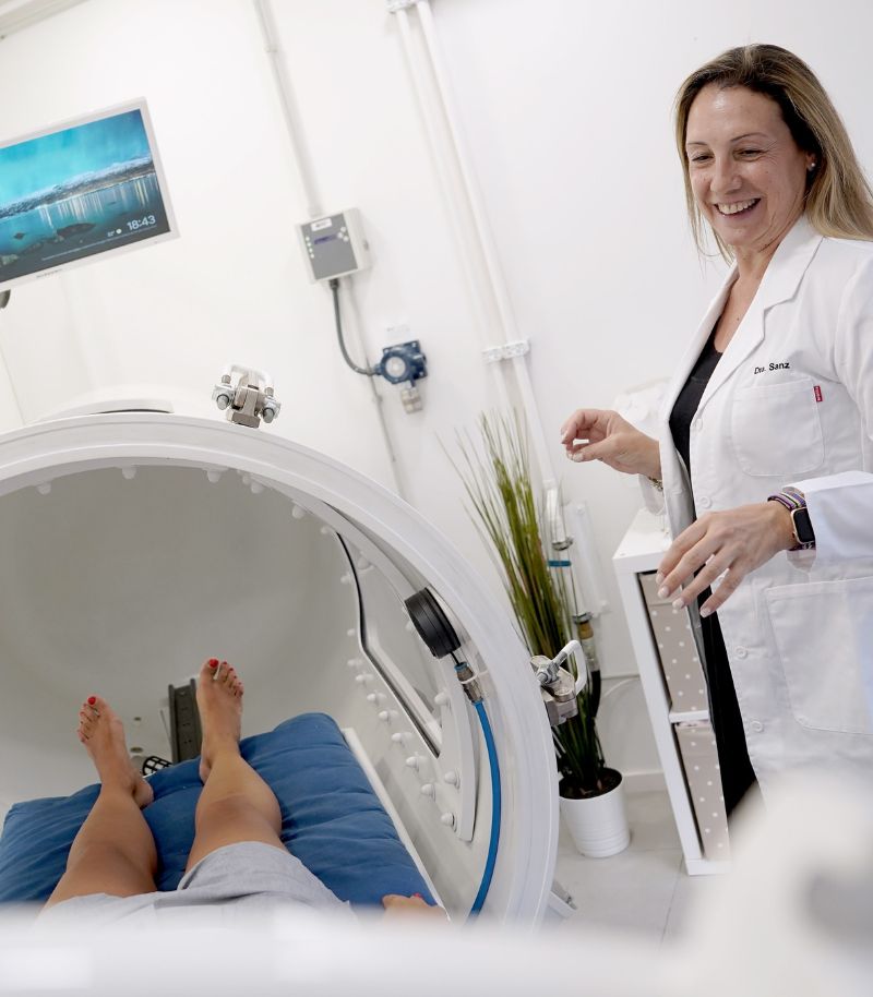
Hyperbaric medicine
Hyperbaric oxygen therapy enhances tissue oxygenation, reduces inflammation and post-surgical oedema, lowers the risk of infection and promotes tissue regeneration and wound healing. It may be considered after basal cell carcinoma surgery, particularly in high-risk patients, following extensive procedures involving grafts or flaps, or in areas with limited vascularisation such as the nose or ears.
Medical and aesthetic treatments
When necessary, specialists in our Facial Aesthetics Unit support the optimisation of surgical outcomes using neuromodulators, dermal fillers or biostimulators to improve skin quality. Laser treatments may also be used to improve scar appearance and achieve optimal functional and aesthetic results. In addition, the Beauty and Wellbeing Unit offers specific post-surgical treatments to promote tissue drainage and accelerate recovery.
Prevention and follow-up
In addition to treating basal cell carcinoma, our unit places strong emphasis on prevention and risk-factor control. Unprotected sun exposure, skin photoageing and certain harmful habits increase the risk of developing new lesions. For this reason, we design personalised prevention and monitoring programmes for each patient.
Preventive laser
Dermatologists in our Laser Unit are involved in research supporting the preventive role of CO₂ rejuvenation laser therapy in the management of the most common skin tumours, including basal cell carcinoma and squamous cell carcinoma.
Ongoing care and personalised follow-up
After treatment for basal cell carcinoma, patients undergo close clinical follow-up and regular check-ups using advanced diagnostic technology, enabling early detection of any abnormalities or recurrences.
What it is, symptoms and treatment
A guide to understanding basal cell carcinoma
This skin tumour originates in the basal cells, which are located at the base of the epidermis, the outermost layer of the skin. These small, round cells play a key role in the renewal of the skin surface, as they produce new cells to replace those that complete their life cycle and are naturally shed.
Basal cell skin cancer most commonly appears in areas of the body that are most exposed to sunlight, such as the face, scalp, ears, shoulders or back, although it may rarely develop in less visible locations. In fact, one of the main risk factors for basal cell carcinoma is prolonged, uncontrolled exposure to ultraviolet radiation.

Risk factors for basal cell carcinoma
In addition to excessive sun exposure, other factors that increase the likelihood of developing basal cell carcinoma include belonging to population groups with lighter skin phototypes (very fair skin, freckles, green, grey or blue eyes, blonde or red hair), having a large number of moles, a family history of skin cancer, frequent sunburns during childhood, excessive or inappropriate use of tanning beds or sun lamps, chronic or recurrent skin damage due to burns, scars or other conditions, as well as spending many hours regularly exposed to the sun without adequate protection (for example, people who work outdoors or spend long periods outdoors practising sports or leisure activities). Men are also more likely to develop basal cell carcinoma than women.
In addition, there are indirect factors that favour the development of basal cell carcinoma. These mainly include smoking, as tobacco damages cellular DNA and increases the overall risk of cancer; having a weakened immune system; previous radiotherapy treatment; prolonged exposure to arsenic; or certain inherited skin disorders, such as xeroderma pigmentosum or basal cell nevus syndrome.
Prognosis and symptoms of basal cell carcinoma
This type of basal cell tumour is more common in patients over the age of 50, mainly due to the cumulative effect of ultraviolet radiation. However, it can develop at any age, particularly in individuals with high levels of sun exposure or a history of frequent sunburns, especially when these occurred during childhood or adolescence.
Prognosis of basal cell carcinoma
Basal cell carcinoma has very limited potential to metastasise and usually grows slowly. Nevertheless, early detection and treatment are essential, as the tumour progresses over time and can become locally invasive (in situ), leading to significant aesthetic damage.
In addition, recurrence after treatment is relatively common. Patients who have previously had basal cell carcinoma should also be aware that this increases the risk of developing other types of skin cancer, such as squamous cell carcinoma.
Symptoms of basal cell carcinoma
The most common sign of basal cell carcinoma is a skin lesion that does not heal — a wound that fails to resolve over time. The appearance of the lesion can vary considerably depending on its location and the patient’s skin phototype.
Basal cell carcinoma may present as a small lump that is skin-coloured or pearly and translucent, often with visible small blood vessels feeding the tumour. This type of lesion is more frequently seen in basal cell carcinomas located on the face and ears. These tumours often bleed and do not heal properly, which is a common reason for patients seeking medical advice.
Basal cell carcinoma lesions may also appear as white, scar-like areas; as darker lesions (brown, black or bluish) with slightly raised, translucent borders and dark spots within; or as reddish, scaly patches with raised edges.
“The risk of metastasis in basal cell carcinoma is very low, but early treatment is essential, as it can be highly locally invasive and lead to significant aesthetic damage”
Treatments for Basal Cell Carcinoma
Skin cancer includes a wide range of neoplasms with different histological characteristics, prognoses and clinical considerations. For this reason, the choice of the most appropriate treatment must always be individualised, taking into account several factors related to:
- The tumour. Including its location, extent, depth of invasion, vascular involvement and whether it has developed on pre-existing skin damage. These factors allow tumours to be classified as low-risk or high-risk.
- The patient’s circumstances. Such as age, life expectancy, clinical condition, risk factors and the importance of aesthetic outcomes.
- The specialist’s judgement. Which depends on experience and expertise not only in skin cancer therapies, but also in the ability to manage potential complications and address functional and aesthetic sequelae. The Grupo Pedro Jaén Skin Cancer Unit is equipped to offer comprehensive care for all types of skin tumours, provided by highly specialised dermatologists with extensive experience in each therapeutic area.
Main treatment options
Curetaje y electrocoagulación
This is a simple and quick technique that generally provides very good results. It is mainly indicated for primary, low-risk basal cell carcinomas, as well as for actinic keratoses (precancerous lesions). The procedure consists of removing the lesion by scraping it with a curette and then applying a high-intensity electrical current to destroy and coagulate the tissue through heat. The result is a wound that must be cared for until complete healing occurs.
In some cases, several sessions may be required, and some patients need local anaesthesia. It should be noted that this treatment can leave scars of variable cosmetic quality, which may make it unsuitable for certain anatomical locations.
Cryotherapy
Cryotherapy is based on freezing the tumour by applying liquid nitrogen in order to destroy cancer cells. The anatomical location must be carefully considered, as this technique causes a controlled freeze injury that, once healed, leaves a scar.
It is suitable for small, superficial primary carcinomas, such as basal cell carcinomas, and can also be used for precancerous lesions such as actinic keratoses. Multiple sessions may be required to achieve complete lesion removal.
Laser treatment
Different types of laser can be used to reduce or destroy tumours, as well as precancerous skin lesions. As with cryotherapy and curettage with electrocautery, laser treatment is indicated for superficial primary carcinomas with a good prognosis. Lasers are also commonly used to treat precancerous and cancerous lesions affecting epithelial linings of organs such as the cervix and the male and female genital area.
Topical treatment
Topical treatment for skin cancer involves medications that modulate and enhance the patient’s immune response. It is mainly used to treat certain precancerous lesions (actinic keratoses) on the face and scalp, as well as small superficial basal cell carcinomas located in visible areas such as the neck, arms, face, décolletage, hands or legs.
The main advantage of this treatment is that it can be applied at home by the patient. The treated area should be washed with soap and water, the cream applied until absorbed, and then left to act for approximately eight hours. For this reason, it is usually recommended to apply it at night before going to bed and to remove any residue and cleanse the skin in the morning.
Patients must avoid direct sun exposure and use high-protection sunscreen (SPF 50). During treatment, a moderate or even intense inflammatory reaction is common, including redness and crust formation. In such cases, the dermatologist will provide specific guidance on how to manage these reactions.
The most common side effects include redness, burning sensation or bleeding in the treated area. As treatment progresses, peeling, dryness, blisters or crusts may appear. Less frequently, patients may experience headache, diarrhoea or fatigue.
These side effects are usually limited to the duration of treatment and resolve once it is discontinued. However, if any side effects occur, patients should consult the prescribing dermatologist to determine the most appropriate course of action.
Surgery with wide margins
Conventional surgery for skin cancer involves surgical excision of the tumour. This procedure is usually performed with widened margins, meaning that a surrounding area of healthy skin is removed as a precaution to ensure complete tumour removal.
The width of the margins depends not only on tumour thickness but also on its location. In small or highly visible areas of the face, it may be difficult or even impossible to remove wide margins due to limited surrounding tissue, and because wider excision results in larger surgical scars.
This approach carries a higher risk of reoperation if the margins obtained in the initial surgery are insufficient to completely remove the tumour.
Mohs surgery
Mohs surgery is a highly effective surgical technique that fulfils three key objectives: complete tumour removal, preservation of function and maximal conservation of healthy tissue. This approach is particularly important when tissue reconstruction is required after tumour excision and is of special relevance given that most skin cancers are located on the face.
This approach involves progressively removing the tumour layer by layer, with each layer examined under a microscope in the operating theatre until no cancer cells are detected. In primary tumours, Mohs surgery achieves cure rates close to 100%, and approximately 98% in recurrent cases. More information about Mohs surgery..
Photodynamic therapy
Photodynamic therapy is a form of photochemotherapy (a therapeutic combination of chemical substances and light) developed to treat certain types of skin cancer lesions in a non-invasive manner.
The treatment consists of applying a photosensitising cream to the affected area and subsequently exposing it to a specific light source, triggering a photochemical reaction that destroys diseased cells without damaging healthy tissue.
Grupo Pedro Jaén was a pioneer in incorporating photodynamic therapy into its Skin Cancer Unit. In addition, some of our specialists have participated in research aimed at developing various applications of photodynamic therapy for both skin tumours and other dermatological conditions.
More information about photodynamic therapy.
Radiotherapy
Radiotherapy is not usually a first-line treatment, but it may be useful in certain situations, such as reducing the size of basal cell carcinomas.
Topical chemotherapy
Topical chemotherapy consists of applying a medication in cream or gel form directly to the lesion being treated. It is used for premalignant lesions and superficial basal cell carcinomas. The advantage of this approach is its highly localised action, which reduces the incidence of systemic side effects.
Nevertheless, the medication may cause burning, itching, erythema (skin redness) and hyperpigmentation. It is generally applied once daily for several weeks, during which the lesion forms a wound that gradually heals until complete resolution.

More information
Below you will find answers to some of the most frequently asked questions about basal cell carcinoma, as well as the authorship and references for the information provided on this page:
Frequently asked questions about basal cell carcinoma
Is basal cell carcinoma dangerous?
Basal cell carcinoma grows very slowly and rarely metastasises. However, it must be treated in its early stages to prevent potentially serious complications, such as infiltration of mucous membranes, nasal cavities or even intracranial structures. In these cases, the prognosis of basal cell carcinoma worsens considerably.
Differences between squamous cell carcinoma and basal cell carcinoma
Basal cell carcinoma originates in the basal cells of the epidermis, whereas squamous cell carcinoma arises from squamous cells. These are the most common types of skin cancer. Although their prognosis is often favourable, they should not be underestimated due to the potential complications they may cause when located in sensitive areas (such as the scalp, nasal cavities or eyelids), either because of tissue loss and associated deformity or their ability to infiltrate vital structures.
Authorship and references
The content of this page has been prepared by the Communication Department and the Medical Team of Grupo Pedro Jaén, in line with our editorial commitment to ensuring the accuracy and up-to-date nature of the information provided.
References on basal cell carcinoma:
1. Kim DP, Kus KJB, Ruiz E. Basal Cell Carcinoma Review. Hematol Oncol Clin North Am. 2019 Feb;33(1):13-24. doi: 10.1016/j.hoc.2018.09.004. PMID: 30497670.
2. Basset-Seguin N, Herms F. Update in the Management of Basal Cell Carcinoma. Acta Derm Venereol. 2020 Jun 3;100(11):adv00140. doi: 10.2340/00015555-3495. PMID: 32346750.
3. Dika E, Scarfì F, Ferracin M, Broseghini E, Marcelli E, Bortolani B, Campione E, Riefolo M, Ricci C, Lambertini M. Basal Cell Carcinoma: A Comprehensive Review. Int J Mol Sci. 2020 Aug 4;21(15):5572. doi: 10.3390/ijms21155572. PMID: 32759706; PMCID: PMC7432343.
4. Marzuka AG, Book SE. Basal cell carcinoma: pathogenesis, epidemiology, clinical features, diagnosis, histopathology, and management. Yale J Biol Med. 2015 Jun 1;88(2):167-79. PMID: 26029015; PMCID: PMC4445438.
5. Tanese K. Diagnosis and Management of Basal Cell Carcinoma. Curr Treat Options Oncol. 2019 Feb 11;20(2):13. doi: 10.1007/s11864-019-0610-0. PMID: 30741348.
6. Di Stefani A, Chimenti S. Basal cell carcinoma: clinical and pathological features. G Ital Dermatol Venereol. 2015 Aug;150(4):385-91. Epub 2015 Jun 23. PMID: 26099353.
7. Dubas LE, Ingraffea A. Nonmelanoma skin cancer. Facial Plast Surg Clin North Am. 2013 Feb;21(1):43-53. doi: 10.1016/j.fsc.2012.10.003. PMID: 23369588.
8. Kauvar AN, Cronin T Jr, Roenigk R, Hruza G, Bennett R; American Society for Dermatologic Surgery. Consensus for nonmelanoma skin cancer treatment: basal cell carcinoma, including a cost analysis of treatment methods. Dermatol Surg. 2015 May;41(5):550-71. doi: 10.1097/DSS.0000000000000296. PMID: 25868035.
9. Peris K, Fargnoli MC, Garbe C, Kaufmann R, Bastholt L, Seguin NB, Bataille V, Marmol VD, Dummer R, Harwood CA, Hauschild A, Höller C, Haedersdal M, Malvehy J, Middleton MR, Morton CA, Nagore E, Stratigos AJ, Szeimies RM, Tagliaferri L, Trakatelli M, Zalaudek I, Eggermont A, Grob JJ; European Dermatology Forum (EDF), the European Association of Dermato-Oncology (EADO) and the European Organization for Research and Treatment of Cancer (EORTC). Diagnosis and treatment of basal cell carcinoma: European consensus-based interdisciplinary guidelines. Eur J Cancer. 2019 Sep;118:10-34. doi: 10.1016/j.ejca.2019.06.003. Epub 2019 Jul 6. PMID: 31288208.
10. Lacour JP. Carcinome basocellulaire [Basal cell carcinoma]. Rev Prat. 1999 Apr 15;49(8):824-8. French. PMID: 10337193.
11. Clark CM, Furniss M, Mackay-Wiggan JM. Basal cell carcinoma: an evidence-based treatment update. Am J Clin Dermatol. 2014 Jul;15(3):197-216. doi: 10.1007/s40257-014-0070-z. PMID: 24733429.
12. Vargo N. Basal cell and squamous cell carcinoma. Semin Oncol Nurs. 2003 Feb;19(1):12-21. doi: 10.1053/sonu.2003.50007. PMID: 12638377.
Where we see patients?
Clinics in Madrid
Grupo Pedro Jaén is a medical group founded more than three decades ago with the aim of providing medical, surgical and aesthetic solutions related to skin health and appearance. Led by Dr Pedro Jaén, the group has a highly specialised medical team that covers every area of care with top-level specialists, carries out extensive training activities, is a leader in medical research, and operates several clinics in Madrid, from which it offers the highest standards of care and clinical excellence to all patients.


Calle Serrano 143
This centre is the main headquarters of Grupo Pedro Jaén. It provides diagnostic and treatment services in skin cancer, clinical and aesthetic dermatology, laser treatments and other related specialties.
Mon - Fri 09:00 - 21:00 / Sat: 09:00 - 14:00
Calle Serrano 143, El Viso neighbourhood, Chamartín district, 28006 Madrid, Spain

La Moraleja
Located in Plaza de La Moraleja, this exclusive centre allows Grupo Pedro Jaén to bring its medical and aesthetic services closer to patients across the entire northern area of Madrid.
Mon - Thu: 09:30 - 20:00 / Fri: 09:30 - 18:00
Calle de la Estafeta 8, ground floor, La Moraleja, 28109 Alcobendas, Madrid
Related units and pages
Skin Cancer
Diagnosis using the most advanced systems, treatment and surgery for the different types of skin cancer.
Mohs Surgery
A surgical technique that allows skin tumours to be removed while preserving as much healthy tissue as possible.
Squamous Cell Carcinoma
The second most common type of skin cancer after basal cell carcinoma, although it is more aggressive.
Actinic Keratosis
A precancerous skin lesion that requires careful monitoring due to its potential to progress.
Skin Cancer Prevention
Based on simple self-care measures and more advanced techniques for early diagnosis.
Clinical dermatology
Highly specialised dermatologists for each skin condition, offering the most advanced treatments available.
Information and news
Press room
In the Grupo Pedro Jaén Press Room you can find information, advice and news related to dermatology, aesthetic medicine, trichology and general health. The group’s journals are also shared through an online platform, along with updates on the activity and media appearances of our clinics and healthcare professionals.

How to shower properly: tips for looking after your skin
Cold or hot water? Is it better to use a sponge? Find out how to shower properly and what dermatologists recommend to protect your skin.
Retinol in summer: should you stop using it?
We debunk this and other myths about using retinol during the warmer months.

Blog
News about Grupo Pedro Jaén. Information and advice on dermatology, aesthetic medicine, trichology, and general health

Media
Information about media appearances by our clinics and medical specialists.

Journal
Archive of all Grupo Pedro Jaén journals, available through an external online platform.