
Skin Cancer Prevention
At our clinics in Madrid: Calle Serrano 143 and La Moraleja
Dermatologists specialising in
Early diagnosis and mole monitoring
Skin cancer is one of the most preventable forms of cancer and also one with the most favourable prognosis when detected at an early stage. For this reason, Grupo Pedro Jaén relies on advanced diagnostic systems and a team of dermatologists specialising in mole monitoring and skin cancer prevention, with a strong focus on early detection and long-term skin health.

Recognised reference clinics in Spain
Home > Skin Cancer > Skin Cancer Prevention
What sets us apart
An innovative approach to active prevention
In addition to clinical monitoring and early diagnosis, Grupo Pedro Jaén incorporates therapeutic strategies with a preventive role in skin cancer. We apply techniques such as photodynamic therapy and fractional CO₂ laser in patients with accumulated sun damage or precancerous lesions, and we actively promote awareness campaigns to reinforce self-care throughout the year. All these measures are integrated into a personalised protocol tailored to each patient’s individual risk profile, supported by advanced technology and multidisciplinary teams with extensive expertise in oncological dermatology.
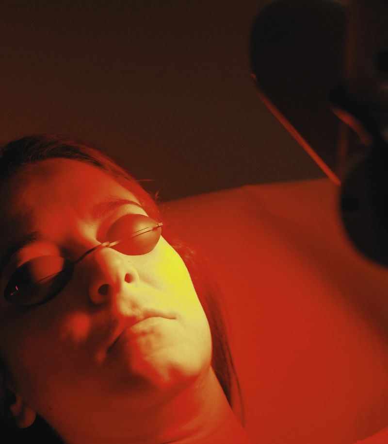
Photodynamic therapy
Photodynamic therapy is a form of non-invasive photochemotherapy developed to treat certain types of skin cancer and precancerous lesions. The treatment combines the application of a topical photosensitising agent with a specific light source, triggering a chemical reaction that selectively destroys damaged cells while preserving healthy surrounding tissue.
Treatment and prevention
Photodynamic therapy is primarily indicated for the treatment of actinic keratoses (precancerous lesions caused by cumulative sun damage) and certain superficial and nodular basal cell carcinomas. Its localised mode of action makes it a safe and effective option for patients with multiple lesions or lesions located in areas where surgery may be more challenging.
Minimal discomfort and rapid recovery
This procedure is quick and does not require general anaesthesia or hospital admission. When necessary, it can also be performed within our Pain-Free Treatments Unit, using local anaesthesia and conscious sedation to minimise discomfort. As a result, photodynamic therapy is a well-tolerated option with a short recovery period.
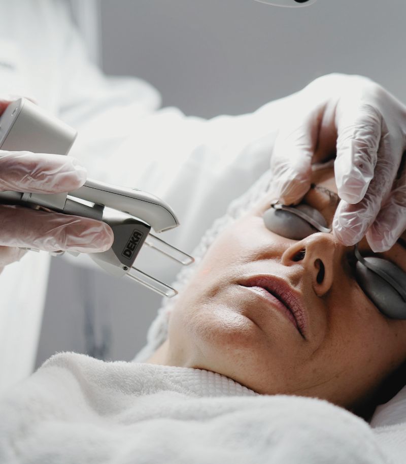
Fractional CO₂ laser
At the Grupo Pedro Jaén Skin Cancer Unit, we closely follow an innovative line of research that is gaining increasing support in international scientific literature: the potential preventive role of fractional CO₂ laser in skin cancer. This device, widely recognised as a gold standard in skin rejuvenation, improves skin quality by removing superficial layers of damaged skin and stimulating deep collagen production.
Promising scientific evidence
In recent years, several studies have suggested that CO₂ laser treatment may reduce the risk of developing actinic keratoses and basal cell carcinoma, the most common type of skin cancer. This effect is thought to be related to the elimination of sun-damaged cells, activation of protective molecular pathways and reduction of senescent cells involved in tumour progression.
Benefits for high-risk patients
This approach is particularly relevant for individuals with advanced photoageing or a high risk of developing malignant skin lesions, and may serve as a complementary strategy in comprehensive skin cancer prevention programmes.
Photoprotection and skin care campaigns
At Grupo Pedro Jaén, we develop awareness and self-care campaigns focused on the prevention of skin cancer and its main risk factors.
Education and early diagnosis initiatives
Through our specialised units in clinical dermatology and skin cancer, we carry out regular campaigns aimed at both the general population and high-risk groups. These initiatives include dermatological check-ups, education on warning signs (such as the ABCDE criteria for melanoma), and practical recommendations to incorporate healthy habits and effective sun protection into daily life.
A year-round commitment
These campaigns are not limited to the summer months. Throughout the year, we promote a culture of continuous skin care through educational talks, patient information materials, and participation in the media and professional forums. Our goal is clear: early detection, reduced incidence of skin cancer, and, above all, empowering patients to care for their skin in a proactive, informed and responsible way.
Mole checks and early diagnosis of skin cancer
A guide to understanding skin cancer prevention
Despite being a common type of cancer and the fact that some of its variants can be life-threatening, skin cancer is one of the most preventable cancers and also one with the most favourable prognosis when detected at an early stage. For this reason, preventive strategies can be broadly divided into two main areas:
- Self-care measures.
- Regular dermatological check-ups and mole monitoring.
Self-care to prevent skin cancer
Almost no disease can be prevented 100% through lifestyle habits or behaviours alone, and skin cancer is no exception. Genetic background, medical history, age or ethnicity are factors that cannot be modified by the patient. However, there are other elements that can be controlled, the most important of which is sun protection.
Sun protection aims to prevent or reduce ultraviolet (UV) radiation–induced DNA damage, which is directly linked to the development of skin cancer. To achieve this, it is essential to:
Use broad-spectrum, high-protection sunscreen
This means applying a broad-spectrum sunscreen that protects against UVA and UVB radiation, as well as infrared radiation, blue light and visible light, with a sun protection factor (SPF) of at least 30. Sunscreen should be reapplied frequently and used every day of the year, even on cloudy days.
Protect children
Dermatologists repeatedly emphasise that the skin has memory and that the consequences of skin cancer often appear decades after cumulative exposure to ultraviolet radiation. For this reason, protecting children from sun damage is essential to reduce their future risk of skin cancer.
In general terms, infants under six months of age should not be exposed to the sun under any circumstances. From that age onwards, sun exposure should be very limited and always accompanied by protective clothing, sunglasses, hats or caps, as well as sunscreen on uncovered areas. During beach or pool activities, children should remain in shaded areas whenever possible. Older children should use highly resistant, age-appropriate sunscreens that must be reapplied frequently, in addition to wearing sunglasses, hats and/or UV-protective clothing when exposed to the sun, particularly at the beach, swimming pool or at sea.
Avoid sunburn
Sunburn causes irreversible DNA damage in skin cells. Experiencing multiple sunburns during childhood and adolescence significantly increases the risk of developing skin cancer in adulthood. In this regard, it is important to stress that there is no such thing as a “healthy tan”: as soon as the skin turns red, it has already begun to burn.
Regular skin self-examination
Examine your skin once a month, paying attention to the appearance of new moles or changes in existing ones, as well as wounds or small ulcers that do not heal properly. Use mirrors or ask for help from another person to examine less accessible areas of the body. Pay particular attention to lesions that are dark brown, black or bluish in colour. Any suspicious change should be assessed by a dermatologist, as these transformations may be early warning signs of skin cancer.

Mole checks for skin cancer prevention
Moles, or melanocytic naevi, are dark, round skin lesions with well-defined borders caused by a localised accumulation of melanocytes. They usually measure between 1 and 6 millimetres in diameter, are flat or slightly raised, and range in colour from light brown to dark brown, café-au-lait tones, black or bluish.
Some babies are born with moles (congenital naevi), but most develop during childhood and adolescence and may appear anywhere on the skin. Nearly everyone has at least one mole, and most people have between 10 and 40. The number of moles is influenced by genetics and cumulative sun exposure over a lifetime.
In most cases, moles are benign. However, many benign and malignant skin lesions can resemble moles. The most common include:
- Seborrhoeic keratoses: Raised, waxy-looking lesions with a rough surface, greyish or brown in colour, typically appearing on the head, neck, back or chest in people over 50.
Solar lentigines: Light brown spots caused by chronic sun exposure, commonly found on the face, shoulders and backs of the hands. - Dermatofibromas: Pigmented lesions with a firmer central area on palpation, usually located on the arms or legs.
- Cherry angiomas: Often referred to as “red moles”, although they are not true moles but vascular lesions visible through the skin.
- Basal cell carcinoma: A very common type of skin cancer that may resemble an intradermal naevus in its early stages.
- Malignant melanoma: A pigmented skin cancer arising from melanocytes. Approximately 75% of melanomas develop on apparently normal skin, while the remaining 25% arise from pre-existing moles.
Understanding these distinctions greatly improves the effectiveness of skin self-examination and early detection of suspicious changes. Dermatologists commonly recommend two practical approaches for mole self-monitoring: the ABCDE rule and the “ugly duckling” sign.
The ABCDE rule
This mnemonic rule helps identify features that may indicate a malignant skin lesion and warrant dermatological assessment.
A – Asymmetry
A mole in which one half does not match the other is suggestive of melanoma.
B – Border
Irregular, poorly defined or uneven borders should raise suspicion.
C – Colour
Most moles are brown to varying degrees, although some may be red, blue or black. Lesions that become very pale, pinkish or even white should be assessed by a dermatologist.
D – Diameter
Although not definitive on its own, moles larger than 6 millimetres require closer observation.
E – Evolution
This is the most important criterion. Any recent change in size, shape, colour, texture or symptoms such as itching, bleeding, oozing or pain should prompt medical evaluation. Evolution refers to any lesion that becomes noticeably different from others on the body or from its previous appearance.
Identifying the “ugly duckling”
Because interpreting the ABCDE rule can be challenging, dermatologists often recommend looking for the so-called “ugly duckling”: a mole that clearly looks different from the rest or from the surrounding skin.
Any lesion that appears unusual or stands out from others should be examined by a dermatologist, even if it does not strictly meet the ABCDE criteria.
Mole monitoring in the dermatology clinic
Although skin self-examination is an important first step, many patients do not examine their skin thoroughly enough to detect warning signs. In addition, these signs may be subtle and easily overlooked.
Some areas of the body are also difficult to examine without assistance. While the arms, hands, face and chest are relatively easy to inspect, this is not the case for the scalp, neck or back.
The Grupo Pedro Jaén Skin Cancer Unit has been a pioneer in implementing systematic mole monitoring to facilitate early detection of skin cancer and to provide the most appropriate treatment for each patient.
Given the association between the number and size of moles and melanoma risk, our specialists recommend an annual dermatological examination. This includes a comprehensive assessment of the entire skin surface, including less visible areas such as the scalp, soles of the feet, spaces between fingers and toes, and the axillae.
The optimal time for mole checks
Spring is generally considered the most suitable time for mole assessment, as the skin is not tanned and lesions retain their natural appearance, making changes easier to detect. During summer, tanning and increased pigmentation can obscure subtle alterations.
However, if any suspicious change is noticed, a dermatology appointment should be scheduled regardless of the time of year.
Ideally, periodic examinations allow for the creation of a personal record of moles, making it possible to monitor the appearance of new lesions, disappearance of others, and any changes over time. In this context, full-body mapping technology, available at our centre, is particularly valuable.
This advanced system captures high-resolution images and generates a complete body map of moles and pigmented lesions, enabling precise monitoring over time and earlier, more accurate detection of clinically significant changes.
“Skin cancer is one of the most preventable cancers and perhaps the one with the most favourable prognosis when detected at an early stage”
Skin cancer diagnosis
Grupo Pedro Jaén has access to the most advanced technologies and procedures for the early detection and accurate classification of skin tumours. This is a key factor in improving patient prognosis and enabling timely, personalised treatment.
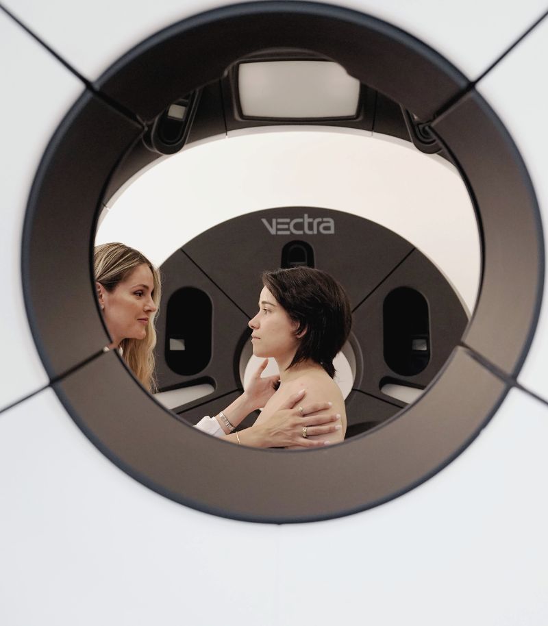
Full-body mapping (Vectra®)
Full-body mapping is currently one of the most accurate technologies for detecting skin cancer, particularly melanoma at its earliest stages. Early detection is directly linked to tumour behaviour and patient survival in melanoma, which, although less common than other skin cancers, is significantly more aggressive and potentially life-threatening.
Grupo Pedro Jaén is currently the only centre in Spain equipped with this technology. The system captures images of the entire skin surface in just two seconds and processes them in three dimensions, providing an exceptionally detailed analysis of every mole or skin lesion within minutes.
The full-body mapping system we use (Vectra® 360) allows rapid, non-invasive examination of the entire skin surface, including areas that may be overlooked during self-examination or are difficult to assess in a standard consultation, such as the groin, axillae, and certain areas of the neck or scalp.
In addition, this technology enables the creation of a personalised dermatological map that records the evolution of moles and skin lesions over time. This makes it easier for specialists to identify suspicious changes quickly and accurately, allowing prompt diagnosis and early treatment.
Virtual biopsy with confocal microscopy
Confocal microscopy represents a major breakthrough in the diagnosis and follow-up of skin lesions. This technology—developed with the active involvement of Dr Salvador González, a member of the Grupo Pedro Jaén team and one of the most internationally recognised skin cancer specialists—offers diagnostic accuracy comparable to that of a conventional biopsy, without the need to remove tissue.
This technique does not require contrast injection, radiation exposure or any invasive procedure. As a result, it significantly reduces the number of traditional biopsies required, providing both physical and emotional benefits for patients. On a physical level, confocal microscopy avoids pain, discomfort and scarring—particularly important when lesions are located on the face or other highly visible areas. On an emotional level, it reduces the anxiety associated with waiting for biopsy results.
The advantages of confocal microscopy extend beyond diagnosis. It is also a valuable tool for monitoring the response to treatment in skin cancer, including non-invasive therapies such as photodynamic therapy, as well as cryotherapy and surgical procedures. This allows specialists to confirm treatment effectiveness or adjust therapeutic strategies when necessary.
Ultrasound
Cutaneous ultrasound plays an increasingly important role in the comprehensive management of skin cancer, particularly in the diagnosis and precise delineation of certain tumours. It is especially useful for lesions that extend deeper into the dermis or subcutaneous tissue, where clinical examination alone may be insufficient.
High-resolution ultrasound provides detailed information about the lesion, including its true size, morphology and depth. This information is essential for assessing tumour behaviour and complements other diagnostic techniques.
In addition, ultrasound helps identify potential involvement of relevant anatomical structures such as blood vessels or nerve endings, which is crucial when planning surgical treatment. This contributes to more accurate and safer preoperative planning, maximising preservation of healthy tissue and optimising oncological, functional and aesthetic outcomes.
Taken together, cutaneous ultrasound has become a highly valuable tool in the integrated management of skin cancer, supporting more informed clinical decision-making and a more personalised approach for each patient.
Digital dermoscopy
The introduction of digital dermoscopy marked a major advance in skin cancer diagnosis, particularly melanoma. This non-invasive technique allows highly magnified imaging of moles, revealing not only surface features but also deeper structural patterns with exceptional detail. Digital dermoscopy is complementary to 3D Total Body Photography (Vectra®) and enables digital monitoring of individual lesions over time.
The technique uses a dermatoscope—a device that combines magnification with specialised lighting to transilluminate the skin and produce high-resolution images. The Vectra® system incorporates the VEOS® dermatoscope, currently considered one of the most precise devices available.
Images captured during each visit are stored and compared over time, allowing accurate assessment of lesion evolution. Digital dermoscopy is quick, painless and completely safe. Its high diagnostic accuracy significantly facilitates early detection of malignant transformation, enabling simpler and more effective surgical treatment when needed. At the same time, it helps avoid unnecessary removal of atypical-looking but benign moles, reducing overtreatment and improving patient care.

More information
Below you will find the authorship and references for the information provided on this page:
Authorship and references
The content of this page has been prepared by the Communication Department and the Medical Team of Grupo Pedro Jaén, in line with our editorial commitment to ensuring the accuracy and up-to-date nature of the information provided.
References on skin cancer prevention:
1. Kornek T, Augustin M. Skin cancer prevention. J Dtsch Dermatol Ges. 2013 Apr;11(4):283-96; quiz 297-8. doi: 10.1111/ddg.12066. PMID: 23574893.
2. Pérez LL, Bashline B. Skin Cancer: Prevention. FP Essent. 2019 Jun;481:28-31. PMID: 31188550.
3. Linos E, Katz KA, Colditz GA. Skin Cancer-The Importance of Prevention. JAMA Intern Med. 2016 Oct 1;176(10):1435-1436. doi: 10.1001/jamainternmed.2016.5008. PMID: 27459394; PMCID: PMC5489348.
4. Lopez AT, Carvajal RD, Geskin L. Secondary Prevention Strategies for Nonmelanoma Skin Cancer. Oncology (Williston Park). 2018 Apr 15;32(4):195-200. PMID: 29684233.
5. Ahmed B, Qadir MI, Ghafoor S. Malignant Melanoma: Skin Cancer-Diagnosis, Prevention, and Treatment. Crit Rev Eukaryot Gene Expr. 2020;30(4):291-297. doi: 10.1615/CritRevEukaryotGeneExpr.2020028454. PMID: 32894659.
6. Dzwierzynski WW. Melanoma Risk Factors and Prevention. Clin Plast Surg. 2021 Oct;48(4):543-550. doi: 10.1016/j.cps.2021.05.001. Epub 2021 Jul 2. PMID: 34503715.
7. American Academy of Dermatology Ad Hoc Task Force for the ABCDEs of Melanoma, Tsao H, Olazagasti JM, Cordoro KM, Brewer JD, Taylor SC, Bordeaux JS, Chren MM, Sober AJ, Tegeler C, Bhushan R, Begolka WS. Early detection of melanoma: reviewing the ABCDEs. J Am Acad Dermatol. 2015 Apr;72(4):717-23. doi: 10.1016/j.jaad.2015.01.025. Epub 2015 Feb 16. PMID: 25698455.
8. Duarte AF, Sousa-Pinto B, Azevedo LF, Barros AM, Puig S, Malvehy J, Haneke E, Correia O. Clinical ABCDE rule for early melanoma detection. Eur J Dermatol. 2021 Dec 1;31(6):771-778. doi: 10.1684/ejd.2021.4171. PMID: 35107069.
9. American Academy of Dermatology Ad Hoc Task Force for the ABCDEs of Melanoma, Tsao H, Olazagasti JM, Cordoro KM, Brewer JD, Taylor SC, Bordeaux JS, Chren MM, Sober AJ, Tegeler C, Bhushan R, Begolka WS. Early detection of melanoma: reviewing the ABCDEs. J Am Acad Dermatol. 2015 Apr;72(4):717-23. doi: 10.1016/j.jaad.2015.01.025. Epub 2015 Feb 16. PMID: 25698455.
10. Stoddard M, Lyons A, Moy R. Skin Cancer Prevention: A Review of Current Oral Options Complementary to Sunscreens. J Drugs Dermatol. 2018 Dec 1;17(12):1266-1271. PMID: 30586257.
11. Brunssen A, Waldmann A, Eisemann N, Katalinic A. Impact of skin cancer screening and secondary prevention campaigns on skin cancer incidence and mortality: A systematic review. J Am Acad Dermatol. 2017 Jan;76(1):129-139.e10. doi: 10.1016/j.jaad.2016.07.045. Epub 2016 Oct 1. PMID: 27707591.
Where we see patients?
Clinics in Madrid
Grupo Pedro Jaén is a medical group founded more than three decades ago with the aim of providing medical, surgical and aesthetic solutions related to skin health and appearance. Led by Dr Pedro Jaén, the group has a highly specialised medical team that covers every area of care with top-level specialists, carries out extensive training activities, is a leader in medical research, and operates several clinics in Madrid, from which it offers the highest standards of care and clinical excellence to all patients.


Calle Serrano 143
This centre is the main headquarters of Grupo Pedro Jaén. It provides diagnostic and treatment services in skin cancer, clinical and aesthetic dermatology, laser treatments and other related specialties.
Mon - Fri 09:00 - 21:00 / Sat: 09:00 - 14:00
Calle Serrano 143, El Viso neighbourhood, Chamartín district, 28006 Madrid, Spain

La Moraleja
Located in Plaza de La Moraleja, this exclusive centre allows Grupo Pedro Jaén to bring its medical and aesthetic services closer to patients across the entire northern area of Madrid.
Mon - Thu: 09:30 - 20:00 / Fri: 09:30 - 18:00
Calle de la Estafeta 8, ground floor, La Moraleja, 28109 Alcobendas, Madrid
Related units and pages
Skin Cancer
Diagnosis using the most advanced systems, treatment and surgery for the different types of skin cancer.
Basal Cell Carcinoma
Early diagnosis and treatment of the most common type of skin cancer, using the most advanced techniques currently available.
Squamous Cell Carcinoma
The second most common type of skin cancer after basal cell carcinoma, although it is more aggressive.
Actinic Keratosis
A precancerous skin lesion that requires careful monitoring due to its potential to progress.
Melanoma
Early diagnosis and treatment of this aggressive skin cancer using the most advanced techniques and technologies.
Clinical dermatology
Highly specialised dermatologists for each skin condition, offering the most advanced treatments available.
Information and news
Press room
In the Grupo Pedro Jaén Press Room you can find information, advice and news related to dermatology, aesthetic medicine, trichology and general health. The group’s journals are also shared through an online platform, along with updates on the activity and media appearances of our clinics and healthcare professionals.

How to shower properly: tips for looking after your skin
Cold or hot water? Is it better to use a sponge? Find out how to shower properly and what dermatologists recommend to protect your skin.

Retinol in summer: should you stop using it?
We debunk this and other myths about using retinol during the warmer months.

Blog
News about Grupo Pedro Jaén. Information and advice on dermatology, aesthetic medicine, trichology, and general health

Media
Information about media appearances by our clinics and medical specialists.

Journal
Archive of all Grupo Pedro Jaén journals, available through an external online platform.