
Confocal microscopy
At our clinics in Madrid: Calle Serrano 143 and La Moraleja
Dermatologists specialising in
Confocal microscopy (virtual skin biopsy)
Confocal microscopy represents a major breakthrough in the diagnosis and follow-up of skin lesions. This technology—developed with the active involvement of Dr Salvador González, a member of the Grupo Pedro Jaén team and one of the most internationally recognised skin cancer specialists—offers diagnostic accuracy comparable to that of a conventional biopsy, without the need to remove tissue.

Recognised reference clinics in Spain
Inicio > Cáncer de piel > Diagnosis > Microscopía confocal
What sets us apart
Diagnostic accuracy without tissue removal
Confocal microscopy enables immediate, real-time diagnosis of skin lesions, supporting more precise and timely clinical decision-making without unnecessary waiting. Its ability to analyse cellular structures at high resolution significantly reduces the need for invasive procedures and helps minimise the removal of healthy tissue. Beyond its role in skin cancer diagnosis, confocal microscopy is also a key tool for monitoring dermatological treatments. It allows repeated, accurate assessment of lesion evolution without the need for multiple biopsies, improving both diagnostic confidence and patient comfort.
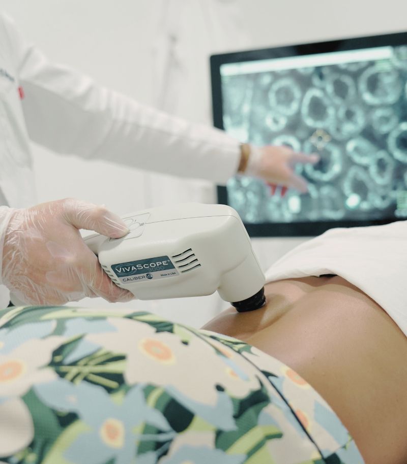
Rapid diagnosis without biopsy
Confocal microscopy has transformed dermatological diagnosis by allowing suspicious skin lesions to be evaluated without the need for a traditional biopsy. By generating real-time images of skin structures at a cellular level, it provides diagnostic accuracy comparable to histopathology while preserving tissue integrity.
Non-invasive and safe technology
This system uses a laser as its light source to examine the skin in vivo. It does not require contrast injections, incisions or exposure to ionising radiation. As a result, it is a safe and well-tolerated technique that reduces patient discomfort and anxiety associated with invasive diagnostic procedures.
Faster clinical decision-making
Real-time results not only eliminate prolonged waiting times but also allow treatment decisions to be made more quickly and safely. This is particularly important in conditions where early intervention is critical, such as melanoma and other skin tumours.
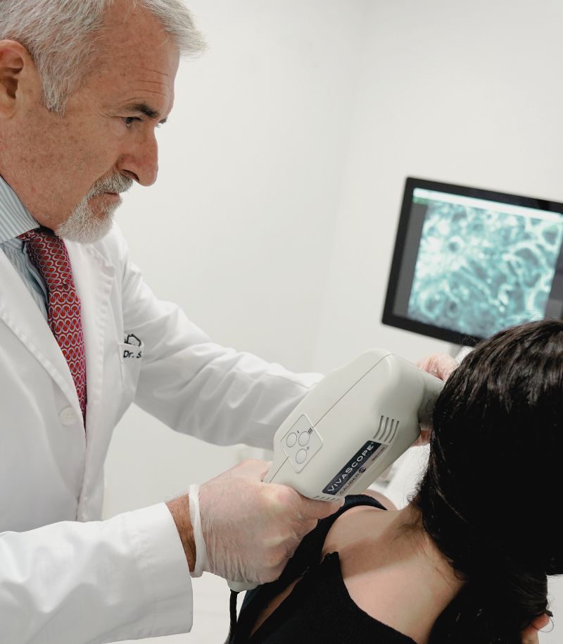
Close monitoring of lesion evolution
Confocal microscopy is not limited to initial diagnosis. It also enables detailed follow-up of skin lesions and assessment of response to different dermatological treatments, allowing therapeutic strategies to be adjusted when necessary.
Real-time assessment of treatment response
This technology makes it possible to evaluate the effectiveness of treatments such as cryotherapy, photodynamic therapy or intralesional treatments before completing the full therapeutic course. In this way, early modifications can be made if the response is suboptimal, helping to prevent persistence or progression of lesions.
Fewer unnecessary surgical procedures
By improving diagnostic certainty, confocal microscopy reduces the number of surgical excisions performed on lesions that later prove to be benign. This is especially relevant for lesions located on the face or other highly visible areas, where avoiding unnecessary tissue removal helps preserve both function and appearance.
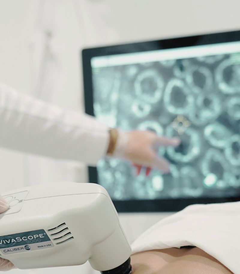
Safer procedures and better cosmetic outcomes
The confocal microscope provides cross-sectional, high-resolution images of the skin, similar to those obtained with advanced imaging techniques, allowing detailed analysis of cellular structures while respecting the integrity of the skin.
Minimising aesthetic impact
In addition to avoiding unnecessary biopsies, confocal microscopy can guide biopsy sampling when tissue removal is unavoidable. This allows more precise targeting of the area of interest, minimising the amount of tissue removed and reducing scarring and post-procedural discomfort.
No side effects
Confocal microscopy is performed by placing a small ring over the lesion, to which the imaging device is attached. Images are obtained without the need for injections, incisions or punctures. As a result, patients avoid pain, scarring and the side effects associated with conventional diagnostic methods.
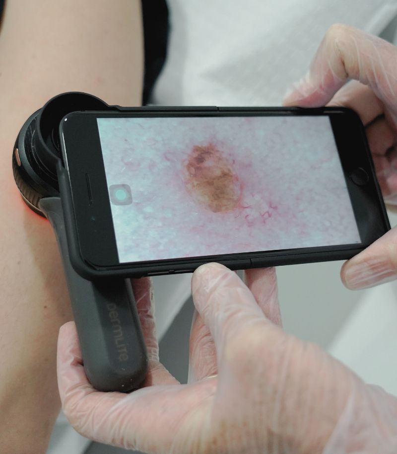
Multiple applications in dermatology
Confocal microscopy plays an important role in the diagnosis and follow-up of a wide range of skin conditions. It provides valuable information for designing effective treatment strategies and for predicting and monitoring therapeutic response.
Skin cancer and melanoma
The combination of confocal microscopy and high-resolution dermoscopy allows accurate pre-surgical diagnosis of melanoma, basal cell carcinoma, squamous cell carcinoma and other skin tumours. This facilitates timely treatment planning, which is essential given the importance of early intervention in skin cancer.
Other dermatological conditions
Confocal microscopy can also be used in the assessment and monitoring of inflammatory skin diseases such as psoriasis, rosacea and hidradenitis suppurativa; blistering disorders (such as pemphigus or epidermolysis bullosa); skin infections; and autoimmune conditions such as vitiligo.
What it involves, effects and recommendations
A comprehensive guide to confocal microscopy
The confocal laser microscope is essentially an optical microscope that uses a laser as its light source and incorporates an electronic system capable of capturing images with exceptional detail and precision.
This imaging technology achieves a much higher resolution than other diagnostic tools and produces extremely accurate images by eliminating interference from light originating in different optical planes throughout the thickness of the observed tissue. As a result, the focus is restricted to a single plane (confocal), significantly improving image clarity.
In clinical practice, confocal microscopy provides cross-sectional images of a specific skin lesion in a way similar to a conventional scanner used in other areas of the body. This allows dermatologists to analyse cells and tissue structures in vivo, in a manner that fully respects skin integrity and without the need for contrast agents, tissue sampling or injections.
With confocal microscopy, it is possible to visualise the margins of proliferative skin lesions in real time, with diagnostic accuracy that is practically equivalent to that of a conventional skin biopsy. This capability is crucial for classifying lesions and moles according to their benign or malignant nature.
In addition to its diagnostic role, confocal microscopy has become an essential tool in dermatology for monitoring the response and evolution of both medical and surgical treatments.

What is a virtual biopsy?
Confocal microscopy is the highest-resolution non-invasive diagnostic technique currently available. It allows a virtual biopsy to be obtained, producing greyscale images comparable to those of a conventional biopsy, without the need to remove tissue, this represents one of the most significant advances in the non-invasive diagnosis of skin tumours and in post-treatment follow-up.
The procedure itself is very straightforward. A small ring is placed over the lesion to be examined, after which the device is attached to this ring and image acquisition begins.
“Confocal microscopy is an advanced technique that allows us to be increasingly less invasive and more precise in the treatment of skin lesions”
Indications for confocal microscopy
Confocal microscopy is a non-invasive and completely safe technique. It does not cause adverse effects and has no known contraindications. Its main indications include the following:
Diagnosis
The combination of confocal microscopy and high-resolution 3D dermoscopy allows for the pre-surgical diagnosis of melanoma, basal cell carcinoma, squamous cell carcinoma and other skin tumours, as well as a wide range of additional dermatological conditions. In addition, it allows us to establish an immediate diagnosis of lesions on the face and other anatomically challenging areas. A clear example is its use to confirm that a lesion is benign and can therefore be removed for aesthetic reasons using laser treatment, rather than scheduling a Mohs micrographic surgery with urgency—a technique for excising skin cancer that cannot be delayed.
Follow-up
Confocal microscopy allows confirmation of a diagnosis following a simple in-clinic evaluation and enables precise monitoring of treatment response. It is particularly useful in assessing therapies such as photodynamic therapy or laser treatment. Using this technology, clinicians can determine in real time whether complete clearance has been achieved or whether additional treatment sessions are required to reach the desired outcome.
Minimising aesthetic impact
The confocal microscope provides cross-sectional, high-resolution images of the skin, similar to those obtained with advanced imaging techniques, allowing detailed analysis of cellular structures while respecting the integrity of the skin.
No side effects
Confocal microscopy is performed by placing a small ring over the lesion, to which the imaging device is attached. Images are obtained without the need for injections, incisions or punctures. As a result, patients avoid pain, scarring and the side effects associated with conventional diagnostic methods.

More information
Below you will find the authorship and references for the information provided on this page:
Authorship and references
The content of this page has been prepared by the Communication Department and the Medical Team of Grupo Pedro Jaén, in line with our editorial commitment to ensuring the accuracy and up-to-date nature of the information provided.
References on confocal microscopy:
1. Waldman RA, Grant-Kels JM. Reflectance confocal microscopy to facilitate knifeless skin cancer management. Cutis. 2019 Oct;104(4):213. PMID: 31774886.
2. Haroon A, Shafi S, Rao BK. Using Reflectance Confocal Microscopy in Skin Cancer Diagnosis. Dermatol Clin. 2017 Oct;35(4):457-464. doi: 10.1016/j.det.2017.06.007. Epub 2017 Aug 7. PMID: 28886802.
3. González S, Sánchez V, González-Rodríguez A, Parrado C, Ullrich M. Confocal microscopy patterns in nonmelanoma skin cancer and clinical applications. Actas Dermosifiliogr. 2014 Jun;105(5):446-58. English, Spanish. doi: 10.1016/j.ad.2013.03.010. Epub 2013 Sep 1. PMID: 24002008.
4. Prow TW, Tan JM, Pellacani G. Reflectance confocal microscopy: hallmarks of keratinocyte cancer and its precursors. Curr Probl Dermatol. 2015;46:85-94. doi: 10.1159/000366541. Epub 2014 Dec 18. PMID: 25561211.
5. Ulrich M, Lange-Asschenfeldt S, González S. In vivo reflectance confocal microscopy for early diagnosis of nonmelanoma skin cancer. Actas Dermosifiliogr. 2012 Nov;103(9):784-9. English, Spanish. doi: 10.1016/j.ad.2011.10.017. Epub 2012 May 15. PMID: 22591654.
6. Ko RF, Smidt AC, Durkin JR. Reflectance confocal microscopy in pediatric dermatology: A state-of-the-art review. Pediatr Dermatol. 2021 Nov;38(6):1488-1499. doi: 10.1111/pde.14837. Epub 2021 Oct 14. PMID: 34651341.
7. Bayguinov PO, Oakley DM, Shih CC, Geanon DJ, Joens MS, Fitzpatrick JAJ. Modern Laser Scanning Confocal Microscopy. Curr Protoc Cytom. 2018 Jul;85(1):e39. doi: 10.1002/cpcy.39. Epub 2018 Jun 20. PMID: 29927100.
8. Elliott AD. Confocal Microscopy: Principles and Modern Practices. Curr Protoc Cytom. 2020 Mar;92(1):e68. doi: 10.1002/cpcy.68. PMID: 31876974; PMCID: PMC6961134.
Where we see patients?
Clinics in Madrid
Grupo Pedro Jaén is a medical group founded more than three decades ago with the aim of providing medical, surgical and aesthetic solutions related to skin health and appearance. Led by Dr Pedro Jaén, the group has a highly specialised medical team that covers every area of care with top-level specialists, carries out extensive training activities, is a leader in medical research, and operates several clinics in Madrid, from which it offers the highest standards of care and clinical excellence to all patients.


Calle Serrano 143
This centre is the main headquarters of Grupo Pedro Jaén. It provides diagnostic and treatment services in skin cancer, clinical and aesthetic dermatology, laser treatments and other related specialties.
Mon - Fri 09:00 - 21:00 / Sat: 09:00 - 14:00
Calle Serrano 143, El Viso neighbourhood, Chamartín district, 28006 Madrid, Spain

La Moraleja
Located in Plaza de La Moraleja, this exclusive centre allows Grupo Pedro Jaén to bring its medical and aesthetic services closer to patients across the entire northern area of Madrid.
Mon - Thu: 09:30 - 20:00 / Fri: 09:30 - 18:00
Calle de la Estafeta 8, ground floor, La Moraleja, 28109 Alcobendas, Madrid
Related units and pages
Skin Cancer Prevention
Based on simple self-care measures and more advanced techniques for early diagnosis.
Skin Cancer
Diagnosis using the most advanced systems, treatment and surgery for the different types of skin cancer.
Melanoma
Early diagnosis and treatment of this aggressive skin cancer using the most advanced techniques and technologies.
Basal Cell Carcinoma
Early diagnosis and treatment of the most common type of skin cancer, using the most advanced techniques currently available.
Squamous Cell Carcinoma
The second most common type of skin cancer after basal cell carcinoma, although it is more aggressive.
Clinical dermatology
Highly specialised dermatologists for each skin condition, offering the most advanced treatments available.
Information and news
Press room
In the Grupo Pedro Jaén Press Room you can find information, advice and news related to dermatology, aesthetic medicine, trichology and general health. The group’s journals are also shared through an online platform, along with updates on the activity and media appearances of our clinics and healthcare professionals.

How to shower properly: tips for looking after your skin
Cold or hot water? Is it better to use a sponge? Find out how to shower properly and what dermatologists recommend to protect your skin.

Retinol in summer: should you stop using it?
We debunk this and other myths about using retinol during the warmer months.

Blog
News about Grupo Pedro Jaén. Information and advice on dermatology, aesthetic medicine, trichology, and general health

Media
Information about media appearances by our clinics and medical specialists.

Journal
Archive of all Grupo Pedro Jaén journals, available through an external online platform.