
Facial Pigmentation
At our centres in Madrid: Serrano 143, Serrano 166 and La Moraleja
Dermatologists specialising in
Treatment of facial hyperpigmentation, including melasma and sunspots
The dermatologists of our Facial Aesthetics Unit have extensive experience in treating different types of facial hyperpigmentation and are supported by our Beauty and Skincare experts, who can prepare depigmentation formulas, and by our Laser Unit for cases where results can be enhanced by using laser technology.

Recognised reference clinics in Spain
Home > Aesthetic Medicine > Facial Aesthetic Medicine > Facial Pigmentation
What sets us apart
Experienced approach with the most advanced therapies
Dark patches on the face represent one of the most common and challenging aesthetic problems. These pigmentation disorders, such as melasma or sunspots, can significantly affect the appearance of the skin, making it look aged and dull. Grupo Pedro Jaén offers a holistic approach to treatment that combines precise dermatological diagnosis, advanced technology and personalised treatments to address each type of dark patch effectively and safely.
Accurate diagnosis
Picosecond laser
Intense pulsed light (IPL)
Magistral formulas
Depigmenting chemical peels
Láser Fraxel
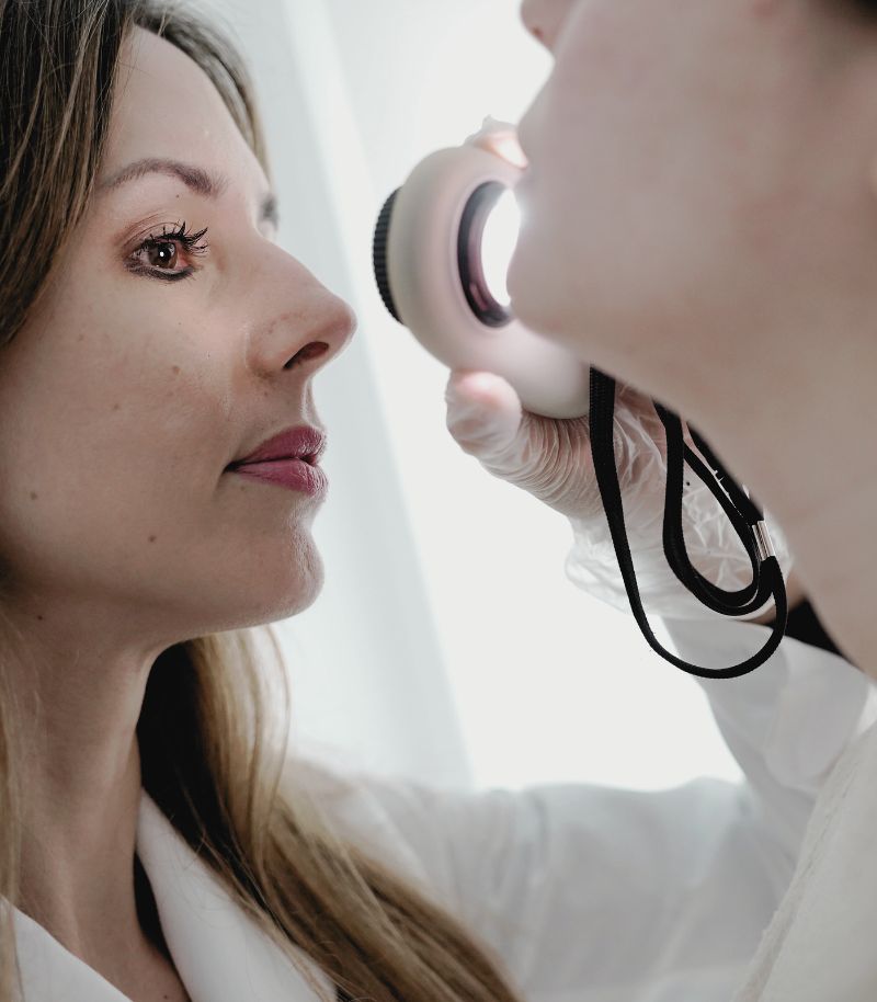
Accurate diagnosis
Before starting any treatment to address pigmented facial skin, it’s essential to understand the type of dark patches, and their cause, to ensure an accurate approach. At Grupo Pedro Jaén, we carry out a detailed dermatological diagnosis that enables us to design a safe and effective treatment, minimising risks such as post-inflammatory hyperpigmentation.
Thorough dermatological evaluation
Our dermatologists who specialise in pigmentary disorders complete a detailed analysis of the skin to differentiate between melasma, sunspots, post-inflammatory hyperpigmentation and guttate hypomelanosis. We use state-of-the-art equipment that enables us to identify the distribution of pigment in the different layers of the skin, and thus tailor the treatment strategy.Multidisciplinary support
Multidisciplinary support
We work in coordination with our Laser Unit and our Beauty and Skincare advisors, combining advanced technology with magistral formulas and continuous dermatological monitoring. In addition, we develop a personalised plan that can include treatments such as picosecond laser, intense pulsed light (IPL) or chemical peels, ensuring optimal results. Through this holistic approach, each patient receives a solution tailored to their specific needs, thus maintaining long-term skin quality and health.
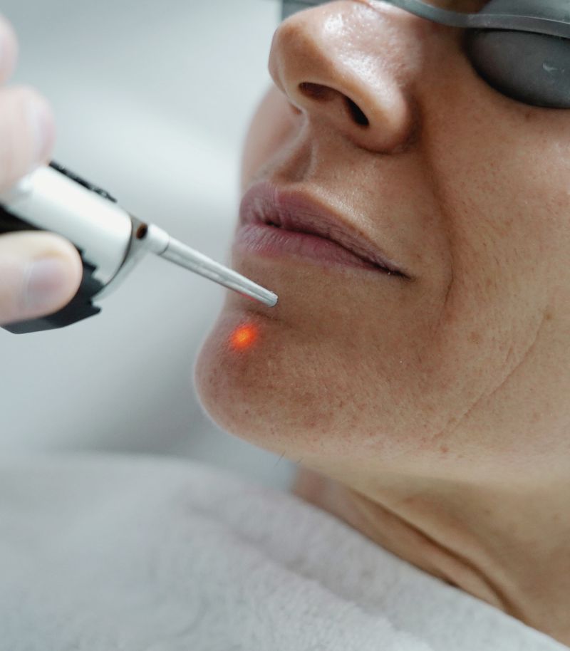
Picosecond laser
The picosecond laser is one of the most advanced technologies for safe and effective removal of dark patches. Its speed in fragmenting melanin enables more efficient removal, reducing the possibility of side effects compared to other types of lasers.
Removal of deep pigmentation
This laser acts on deep pigment deposits, breaking them down into microscopic particles that the body can eliminate naturally. It is especially useful for stubborn dark patches such as dermal melasma, which does not always respond well to other depigmenting treatments.
Precision and visible results in just a few sessions
Thanks to its precision, the picosecond laser minimises the risk of inflammation and provides results in fewer sessions than other treatments. In addition, its rapid action improves the uniformity of skin tone and luminosity without damaging the surrounding tissues, making recovery faster and safer.
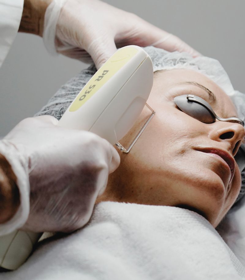
Intense pulsed light (IPL)
Intense pulsed light (IPL) is a versatile option for treating dark patches on the face, neck and hands. Its ability to work at different depths enables multiple pigmentation concerns to be addressed at the same time, offering comprehensive rejuvenation.
Evening out skin tone
This treatment uses pulses of light that fragment the melanin in superficial dark patches, allowing them to be progressively eliminated by the body. It is especially useful for patients with uneven pigmentation due to sun damage, as it not only lightens spots but also improves skin texture.
Complete rejuvenation
When combined with picosecond lasers or depigmenting formulas, IPL boosts results for more even, luminous skin. In addition, its effect on collagen improves the firmness and elasticity of the skin, helping to prevent the appearance of new dark patches.

Magistral formulas
The treatment of facial dark patches requires an approach tailored to the individual patient. At the Grupo Pedro Jaén, we prepare magistral formulas that combine highly effective active ingredients to treat hyperpigmentation safely and effectively.
Treatments tailored to each patient
We develop personalised depigmenting creams, adjusting each formula according to the patient’s skin type, the depth of the pigmentation and skin sensitivity, ensuring effective treatment without adverse effects.
Perfect complement to clinical therapies
These formulas enhance the effects of other treatments such as laser, IPL or chemical peels, helping to maintain results in the long term. Regular use of these creams helps to control pigmentation and prevent the reappearance of dark patches, reinforcing the skin’s protective barrier to keep it in optimal condition.
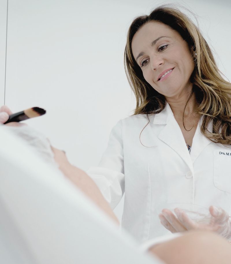
Depigmenting chemical peels
Chemical peels are an effective technique for treating facial hyperpigmentation in a controlled manner. By means of chemical exfoliation, we eliminate pigmented cells and promote cell renewal, obtaining a more luminous and uniform skin.
Controlled exfoliation
Our experts apply acid peels to the face, formulated as appropriate for the type of dark patch and the skin characteristics. These acids renew the surface layer of the skin, accelerating regeneration and facilitating the elimination of accumulated pigmentation.
Progressive and safe results
The results start to become visible after the first session, although in some cases several applications are required to achieve total depigmentation. As it is an adaptable technique, the risk of irritation is minimal when the type of peel is correctly personalised.
Fraxel laser
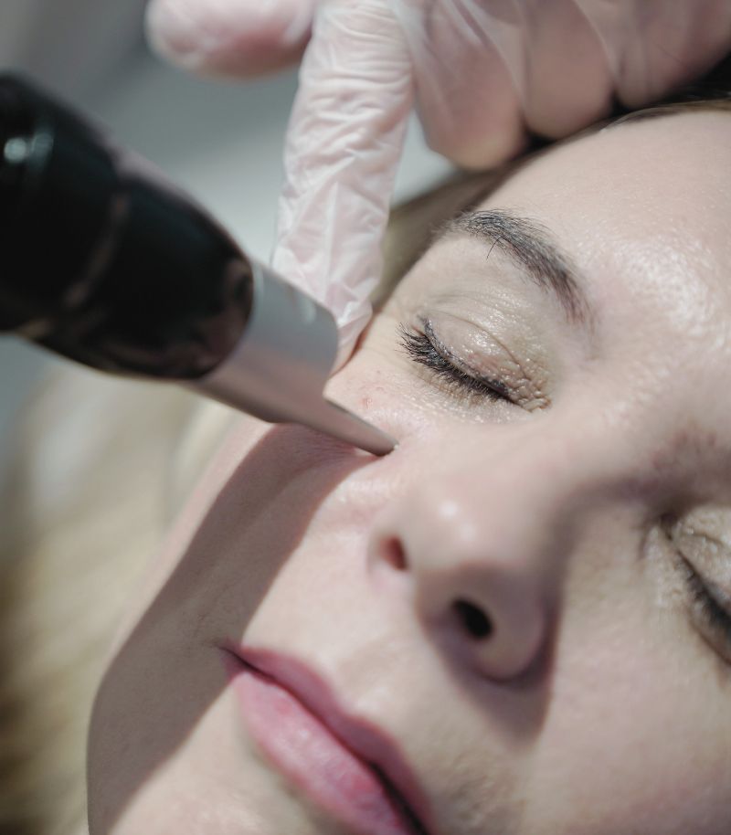
The Fraxel laser is a particularly effective technology for treating dark patches on the skin. It works by creating microperforations in the skin through which the energy passes. This enables areas of tissue to be left intact, which speeds up recovery.
How it treats dark patches
This laser acts deep in the dermis, stimulating collagen production and promoting cell regeneration. This improves the texture and elasticity of the skin, while evening out the skin tone and eliminating hyperpigmented patches. Its action enables it to treat not only superficial pigmentation but also deeper problems, such as fine wrinkles or acne scars.
Additional benefits
The Fraxel laser also improves the luminosity and tone of the skin, giving it a fresher, rejuvenated appearance. The session is quick and side effects are minimal, mainly temporary redness. The results become progressively more evident after several sessions.

Differential diagnosis of melasma
Melasma is a skin condition characterised by dark patches, usually on the face, especially on the cheeks, forehead and upper lip. It is more common in women, is related to hormonal factors and becomes more pronounced with sun exposure.
Ensuring melasma is not aggravated further
Correct diagnosis of melasma is important as it can be confused with other pigmented lesions such as sunspots. Treating melasma incorrectly, for example using lasers to remove it without proper diagnosis and a prior depigmentation treatment, can make the situation worse, causing the patches to become even darker.
Treatment of melasma
As lasers can irritate the melanin and cause an increase in pigmentation, they are not used as the standard treatment and are only applied in specific cases and always after the patient has followed a topical depigmentation treatment at home. It is therefore crucial that a dermatologist or specialised professional makes an accurate diagnosis to determine the type of hyperpigmentation and the appropriate treatment.
What it is, types and treatment
A guide to understanding facial pigmentation
Facial pigmentation disorders are among the skin alterations with the greatest negative impact on facial appearance. These changes in pigmentation affect a large proportion of the population from a certain age onwards and, according to patients themselves, give the face a dull and aged appearance that is often more pronounced than that caused by other aesthetic concerns such as wrinkles or skin laxity. Genetic predisposition, hormonal factors and excessive sun exposure are among the main causes of facial pigmentation. For this reason, these conditions should be assessed and treated by dermatologists with expertise in pigmentary disorders in order to achieve optimal and safe results.
Accurate diagnosis and correct classification of the different types of facial pigmentation are essential to establish an effective treatment strategy. Among the most relevant facial pigmentary disorders are melasma, solar lentigines and idiopathic guttate hypomelanosis.

Solar lentigines
Solar lentigines are brown macules commonly described by patients as “large freckles”. They represent one of the most visible manifestations of photoageing, caused by cumulative damage from prolonged and excessive exposure to ultraviolet radiation.
These lesions have an irregular shape and uneven pigmentation, although always within shades of brown. They most commonly appear on the shoulders, arms, hands, face and décolletage, but may develop on any area of the body that has been chronically exposed to sunlight. In fact, solar lentigines often represent the long-term visible consequence of sunburns experienced during childhood or adolescence.
Solar lentigines tend to appear in clusters and gradually increase in size with age. They are asymptomatic and benign; however, periodic dermatological follow-up is essential to differentiate them from malignant lentigines, which are cancerous lesions that require surgical excision.
Treatment for solar lentigines
Laser treatment is the most effective option for the management of solar lentigines, as these lesions primarily affect the most superficial layers of the skin. Laser therapies work by selectively targeting and destroying the excess melanin responsible for pigmentation
The choice of laser or light-based technology depends on several factors, including the presence of other pigmentary disorders, lesion depth, the number of lentigines to be treated and their anatomical location.
Picosecond alexandrite laser
This is one of the most effective laser technologies for the treatment of a wide range of pigmentary lesions. It is well tolerated, quick to perform and delivers excellent results in a small number of sessions. A key advantage of this laser is its ability to treat deeper pigmentation and hormonal pigmentation disorders, in addition to solar lentigines.
Q-switched laser
Indicated for the treatment of solar lentigines, this laser achieves visible results after only a few sessions.
Optimised intense pulsed light (IPL)
IPL allows treatment of large areas affected by multiple or extensive lentigines. In addition, its effects can be used to simultaneously improve vascular lesions during the same treatment session.
After laser treatment, it is common to experience temporary skin irritation, mild swelling and redness. Pigmented lesions typically darken and form superficial crusts, which may persist for one to three weeks. Once these crusts shed, a faint residual pigmentation may remain temporarily and usually resolves within a few days.
“In most cases, the best results are achieved by combining different therapeutic options and acting at an early stage.”
Melasma
The term melasma derives from the Greek word melas, meaning black. In dermatology, melasma refers to a characteristic form of facial hyperpigmentation with brownish patches, typically symmetrically distributed. Melasma is caused by a combination of genetic and hormonal factors—such as pregnancy, oral contraceptive use and thyroid disorders—and is exacerbated by sun exposure. Lesions are located in sun-exposed areas of the face, tend to worsen during spring and summer, and often improve during autumn and winter.
Melasma can affect both men and women of any age and ethnic background, although it is more common in women of Hispanic origin and in individuals living in tropical regions. While melasma in men is less frequent, it presents similar clinical features and shows a strong familial component, observed in up to 70% of cases.
Hormonal changes during pregnancy are one of the most common triggers. Although pregnancy-related melasma may improve after childbirth, these lesions are often difficult to treat, making prevention and specialist dermatological management particularly important.
Classification of melasma
En función de la profundidad de la hiperpigmentación, se suele hablar de melasma dérmico (afecta a un nivel profundo de la dermis y la coloración de las manchas tiende al grisáceo), melasma epidérmico (se localiza a un nivel más superficial de la piel y las manchas son de color marrón con bordes bien definidos) y melasma mixto (combinación de ambos, caracterizado por manchas pardas y grisáceas).
Based on the depth of pigmentation, melasma is classified as:
- Centrofacial pattern The most common form, involving the cheeks, forehead, upper lip and chinPatrón centro facial.
- Malar pattern, affecting the cheekbones, cheeks and nose
- Mandibular pattern, the least frequent, limited to the jawline
Treatment of melasma
Melasma treatment is complex and requires a carefully planned combination of different therapeutic approaches. It differs significantly from the treatment of solar lentigines, and prognosis depends directly on correct diagnosis and management.
Topical depigmenting agents
These constitute the first-line treatment for melasma. They include lotions and creams containing activeaingredientsathat inhibit melanin production. Exfoliating agents may be added to enhance skin renewal, along with antioxidants to reduce free radical damage. These products may be commercially available or prepared as personalised magistral formulas prescribed by a dermatologist.
Chemical peels
Chemical peels are considered a second-line treatment and are used as an adjunct to topical therapy. Applied in the dermatologist’s office, they help lighten pigmentation by promoting controlled exfoliation and skin renewal.
Laser treatment
Laser treatment is also considered a second-line option and should always be used as a complementary therapy, never as a first or sole treatment for melasma.
Lasers used in melasma management operate at low energy settings to remove pigment without causing skin injury or crust formation, thereby minimising the risk of post-inflammatory hyperpigmentation.
For this reason, it is essential that laser treatment for melasma is performed by an experienced dermatologist with in-depth knowledge of both the condition and laser–skin interactions. Incorrect laser use may lead to rebound hyperpigmentation and worsening of existing lesions.
General measures for melasma management
Independientemente de cuál sea el tratamiento pautado por el dermatólogo experto en melasma. Es necesario que los pacientes sigan una serie de consejos para potenciar la terapia y evitar que empeore.
Photoprotection
Strict sun protection is essential. Broad-spectrum sunscreens with SPF 50+ and physical protection measures—such as sunglasses, wide-brimmed hats and protective clothing—must be used before, during and after treatment. Without adequate photoprotection, melasma treatment will not be effective.
Cosmetic camouflage
Specialised makeup products may be used to conceal localised pigmentation and serve as a complementary measure alongside medical treatments.
Avoidance of irritant or allergenic agents
Certain substances may cause delayed hypersensitivity reactions, leading to cosmetic complications ranging from post-inflammatory hyperpigmentation to irreversible depigmentation.
Therapeutic synergy
At Grupo Pedro Jaén, clinical experience has shown that combining different treatment modalities produces better outcomes than relying on a single approach. Ongoing follow-up is essential to assess response and determine the need for additional laser sessions or adjustments in topical and peeling protocols.

Idiopathic guttate hypomelanosis
Idiopathic guttate hypomelanosis deserves special mention among facial pigmentation disorders. Although it is related to chronic sun exposure, it presents not as dark pigmentation but as white macules.
This condition is characterised by small, round or oval white spots that appear predominantly on sun-exposed areas, especially the arms and legs. These lesions become more noticeable during and after summer due to contrast with surrounding tanned skin.
The condition results from exhaustion of melanocytes, which lose their ability to produce melanin after years of excessive sun exposure. Instead of hyperpigmentation, these areas undergo depigmentation due to impaired melanocyte function.
Treatment of idiopathic guttate hypomelanosis
Although these lesions are clinically asymptomatic, they may be aesthetically concerning. When treatment is desired, the most effective approach currently involves a combination of laser therapy and controlled ultraviolet exposure.
The aim is to stimulate depleted melanocytes to resume melanin production through fractional laser treatment—ablative or non-ablative, depending on clinical judgement—combined with controlled UV exposure.
Treatment typically requires a variable number of sessions to achieve improvement. Strict photoprotection measures must be followed to prevent progression of the condition and to enhance the effectiveness of repigmentation therapy.
More information
Below you will find the authorship and references for the information provided on this page:
Authorship of this content
The content of this page has been prepared by the Communication Department and the Medical Team of Grupo Pedro Jaén, in line with our editorial commitment to ensuring the accuracy and up-to-date nature of the information provided.
References on facial pigmentation:
1. McKesey J, Tovar-Garza A, Pandya AG. Melasma Treatment: An Evidence-Based Review. Am J Clin Dermatol. 2020 Apr;21(2):173-225. doi: 10.1007/s40257-019-00488-w. PMID: 31802394.
2. Spierings NMK. Melasma: A critical analysis of clinical trials investigating treatment modalities published in the past 10 years. J Cosmet Dermatol. 2020 Jun;19(6):1284-1289. doi: 10.1111/jocd.13182. Epub 2019 Oct 11. PMID: 31603285.
3. Rivas S, Pandya AG. Treatment of melasma with topical agents, peels and lasers: an evidence-based review. Am J Clin Dermatol. 2013 Oct;14(5):359-76. doi: 10.1007/s40257-013-0038-4. PMID: 23881551.
4. Bala HR, Lee S, Wong C, Pandya AG, Rodrigues M. Oral Tranexamic Acid for the Treatment of Melasma: A Review. Dermatol Surg. 2018 Jun;44(6):814-825. doi: 10.1097/DSS.0000000000001518. PMID: 29677015.
5. Artzi O, Horovitz T, Bar-Ilan E, Shehadeh W, Koren A, Zusmanovitch L, Mehrabi JN, Salameh F, Isman Nelkenbaum G, Zur E, Sprecher E, Mashiah J. The pathogenesis of melasma and implications for treatment. J Cosmet Dermatol. 2021 Nov;20(11):3432-3445. doi: 10.1111/jocd.14382. Epub 2021 Aug 19. PMID: 34411403.
6. Neagu N, Conforti C, Agozzino M, Marangi GF, Morariu SH, Pellacani G, Persichetti P, Piccolo D, Segreto F, Zalaudek I, Dianzani C. Melasma treatment: a systematic review. J Dermatolog Treat. 2022 Jun;33(4):1816-1837. doi: 10.1080/09546634.2021.1914313. Epub 2022 Mar 23. PMID: 33849384.
7. Passeron T, Picardo M. Melasma, a photoaging disorder. Pigment Cell Melanoma Res. 2018 Jul;31(4):461-465. doi: 10.1111/pcmr.12684. Epub 2018 Jan 12. PMID: 29285880.
8. Borelli C, Fischer S. Chemical Peelings zur Behandlung von Melasma, Pigmentstörungen und Hyperpigmentierungen : Indikationen, Effektivität und Risiken [Chemical peeling for treatment of Melasma, pigmentary disorders and hyperpigmentation : Indications, effectivity and risks]. Hautarzt. 2020 Dec;71(12):950-959. German. doi: 10.1007/s00105-020-04712-1. PMID: 33141276.
9. Plensdorf S, Livieratos M, Dada N. Pigmentation Disorders: Diagnosis and Management. Am Fam Physician. 2017 Dec 15;96(12):797-804. PMID: 29431372.
10. Searle T, Al-Niaimi F, Ali FR. The top 10 cosmeceuticals for facial hyperpigmentation. Dermatol Ther. 2020 Nov;33(6):e14095. doi: 10.1111/dth.14095. Epub 2020 Sep 4. PMID: 32720446.
11. Kerns ML, Chien AL, Kang S. A Role for NRF2-Signaling in the Treatment and Prevention of Solar Lentigines. Plast Reconstr Surg. 2021 Dec 1;148(6S):27S-31S. doi: 10.1097/PRS.0000000000008783. PMID: 34847095.
12. Szymańczyk J, Trzeciakowski W, Ivonyak Y, Tuchowski P, Bercha A, Szymańczyk J. Blue Laser (450 nm) Treatment of Solar Lentigines. J Clin Med. 2021 Oct 24;10(21):4919. doi: 10.3390/jcm10214919. PMID: 34768439; PMCID: PMC8584462.
13. Praetorius C, Sturm RA, Steingrimsson E. Sun-induced freckling: ephelides and solar lentigines. Pigment Cell Melanoma Res. 2014 May;27(3):339-50. doi: 10.1111/pcmr.12232. Epub 2014 Mar 3. PMID: 24517859.
14. Bastiaens M, Hoefnagel J, Westendorp R, Vermeer BJ, Bouwes Bavinck JN. Solar lentigines are strongly related to sun exposure in contrast to ephelides. Pigment Cell Res. 2004 Jun;17(3):225-9. doi: 10.1111/j.1600-0749.2004.00131.x. PMID: 15140067.
15. Araghi F, Ohadi L, Moravvej H, Amani M, Allameh F, Dadkhahfar S. Laser treatment of benign melanocytic lesion: a review. Lasers Med Sci. 2022 Sep 12. doi: 10.1007/s10103-022-03642-9. Epub ahead of print. PMID: 36097230.
16. Brown F, Crane JS. Idiopathic Guttate Hypomelanosis. 2022 May 8. In: StatPearls [Internet]. Treasure Island (FL): StatPearls Publishing; 2022 Jan–. PMID: 29489254.
17. Buch J, Patil A, Kroumpouzos G, Kassir M, Galadari H, Gold MH, Goldman MP, Grabbe S, Goldust M. Idiopathic guttate hypomelanosis: Presentation and Management. J Cosmet Laser Ther. 2021 Feb 17;23(1-2):8-15. doi: 10.1080/14764172.2021.1957116. Epub 2021 Jul 25. PMID: 34304679.
18. Juntongjin P, Laosakul K. Idiopathic Guttate Hypomelanosis: A Review of its Etiology, Pathogenesis, Findings, and Treatments. Am J Clin Dermatol. 2016 Aug;17(4):403-11. doi: 10.1007/s40257-016-0195-3. PMID: 27206417.
Where we see patients?
Clinics in Madrid
Grupo Pedro Jaén is a medical group founded more than three decades ago with the aim of providing medical, surgical and aesthetic solutions related to skin health and appearance. Led by Dr Pedro Jaén, the group has a highly specialised medical team that covers every area of care with top-level specialists, carries out extensive training activities, is a leader in medical research, and operates several clinics in Madrid, from which it offers the highest standards of care and clinical excellence to all patients.

Calle Serrano 143
This centre is the main headquarters of Grupo Pedro Jaén. It provides diagnostic and treatment services in skin cancer, clinical and aesthetic dermatology, laser treatments and other related specialties.
Mon - Fri: 09:00 - 21:00 / Sat: 09:00 - 14:00
Calle Serrano 143, El Viso neighbourhood, Chamartín district, 28006 Madrid, Spain

La Moraleja
Located in Plaza de La Moraleja, this exclusive centre allows Grupo Pedro Jaén to bring its medical and aesthetic services closer to patients across the entire northern area of Madrid.
Mon - Thu: 09:30 - 20:00 / Fri: 09:30 - 18:00
Calle de la Estafeta 8, ground floor, La Moraleja, 28109 Alcobendas, Madrid, Spain

Calle Serrano 166
This centre currently offers services in body aesthetic medicine, endocrinology, nutrition, complementary aesthetic medicine and post-surgical aesthetic treatments.
Mon - Fri: 09:00 - 21:00 / Sat: 09:00 - 14:00
Calle Serrano 166, El Viso neighbourhood, Chamartín district, 28002 Madrid

Related units and pages
Facial aesthetics
Minimally invasive treatments to improve facial appearance with harmonious and natural-looking results.
Body aesthetics
A personalised approach combining different treatment strategies to address the main body-related concerns.
Male aesthetics
Surgical and aesthetic treatments fully adapted to male facial and body anatomy.
Scars
Fully personalised treatments tailored to the type and specific characteristics of each scar.
Plastic surgery
Treatment of facial and body aesthetic and functional concerns that require a surgical solution.
Complementary aesthetics
Cosmetic and aesthetic treatments that address beauty from a clinical perspective to achieve optimal results.
Information and news
Press room
In the Grupo Pedro Jaén Press Room you can find information, advice and news related to dermatology, aesthetic medicine, trichology and general health. The group’s journals are also shared through an online platform, along with updates on the activity and media appearances of our clinics and healthcare professionals.

How to shower properly: tips for looking after your skin
Cold or hot water? Is it better to use a sponge? Find out how to shower properly and what dermatologists recommend to protect your skin.
Retinol in summer: should you stop using it?
We debunk this and other myths about using retinol during the warmer months.

Blog
News about Grupo Pedro Jaén. Information and advice on dermatology, aesthetic medicine, trichology, and general health

Media
Information about media appearances by our clinics and medical specialists.

Journal
Archive of all Grupo Pedro Jaén journals, available through an external online platform.